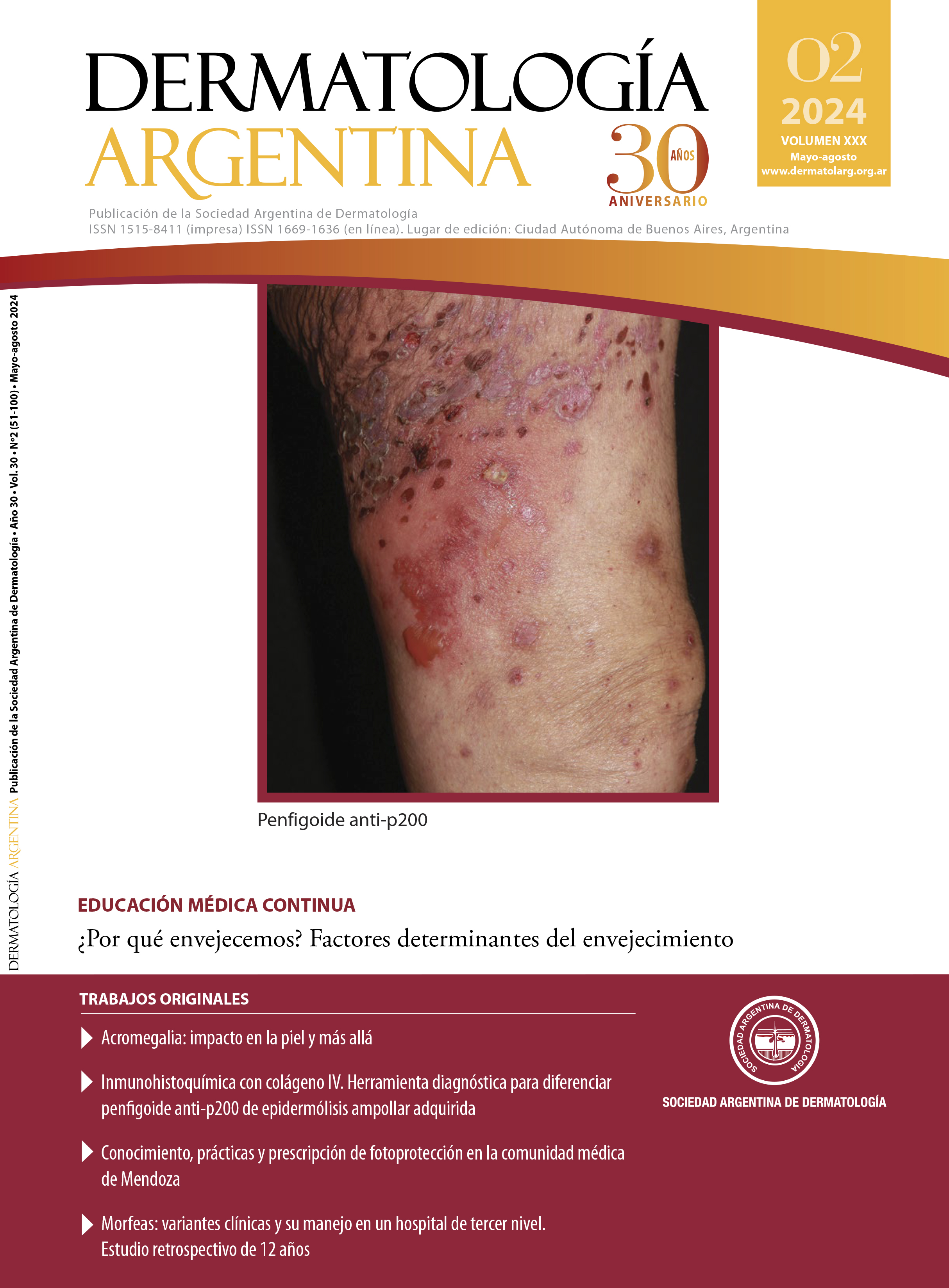
Immunohistochemical with type IV collagen. A diagnostic tool to differentiate anti-p200 pemphigoid from epidermolysis bullosa acquisita
DOI:
https://doi.org/10.47196/da.v30i2.2576Keywords:
immunohistochemical collagen IV, anti-p200 pemphigoid, laminin gamma 1, acquired bullous epidermolysis, autoimmune bullous dermatosesAbstract
Introduction: the diagnosis of autoimmune bullous diseases represents quite a challenge. When the immunofluorescence with salt-split technique has IgG and C3 at the floor of the membrane zone, several differential diagnoses are considered. Immunohistochemical with collagen IV is suggested as a complementary tool to differentiate between acquired bullous epidermolysis and anti-p200 pemphigoid.
Objectives: use immunohistochemical with collagen IV to differentiate anti-p200 pemphigoid from epidermolysis bullosa acquisita. Secondly, describe the epidemiological, clinical, and histopathological characteristics of these patients with these entities within our population.
Design: cross-sectional, retrospective, descriptive, observational, multicenter study.
Materials and methods: immunohistochemical with collagen IV was performed on biopsy specimens from patients whose direct immunofluorescence with salt-split technique showed IgG and C3 deposits on the floor of the membrane zone.
Results: 19 samples were analyzed, 12 (63.2%) testing positive for immunohistochemical with collagen IV, and 7 (36.8%) showing no labeling. Among the positive samples, 8 (42.1%) exhibited labeling on the floor of the membrane zone, interpreted as probable anti-p200 pemphigoid, and 4 (21.1%) on the roof, interpreted as acquired bullous epidermolysis.
Conclusions: immunohistochemical with collagen IV turned out to be an accessible and highly useful method that complements the diagnostic methods of autoimmune bullous dermatoses of the dermoepidermal junction. In this study, we observed a high predominance of cases with a probable diagnosis of anti-p200 pemphigoid, which reinforces the fact that this pathology is underdiagnosed due to the lack of accurate diagnostic methods.
References
I. Maronna E. Histopatología. En: Forero O, Candiz M.E, Olivares L. Dermatosis ampollares autoinmunes. Haga su diagnóstico. Ed Journal. Buenos Aires 2021;10-28
II. Forero O, Roquel L. Inmunofluorescencia. En: Forero O, Candiz M.E, Olivares L. Dermatosis ampollares autoinmunes. Haga su diagnóstico. Ed Journal. Buenos Aires 2021;29-40.
III. Candiz ME. Serologías por ELISA. En: Forero O, Candiz M.E, Olivares L. Dermatosis ampollares autoinmunes. Haga su diagnóstico. Ed Journal. Buenos Aires 2021;41-50.
IV. García-Díez I, Martínez-Escala M.E, Ishii N, Hashimoto, et ál. Descripción de dos casos de penfigoide anti-p200. Utilidad de una técnica inmunohistoquímica sencilla en el diagnóstico diferencial con otras enfermedades ampollosas autoinmunes. Actas Dermosifilogr. 2017;108:e1-e5.
V. Sajeela-Rasheed V. Anti-p200 pemphigoid: a review. J Skin Sex Transm Dis. 2023;5:22-27.
VI. Luzar B, McGrath J. Inherited and autoimmune subepidermal blistering diseases. En: Calonje E, Brenn T, Lazar A, Billings S. McKee’s pathology of the skin with clinical correlations. Elsevier, Edinburgh, 2020:118-170.
VII. Meijer JM, Diercks GF, Schmidt E, Pas HH, et ál. Laboratory diagnosis and clinical profile of anti-p200 pemphigoid. JAMA Dermatol. 2016;152:897-904.
VIII. Ginzburg K, Forero O, Candiz ME, Maronna E, et ál. Penfigoide anti-p200: ¿enfermedad poco frecuente o subdiagnosticada? Dermatol Argent. 2022;28:25-29.
IX. Forero O, Candiz ME. Epidermólisis ampollar adquirida, variedad inflamatoria símil penfigoide de las mucosas. En: Forero O, Candiz M.E, Olivares L. Dermatosis ampollares autoinmunes. Haga su diagnóstico. Ed Journal. Buenos Aires 2021; 231-234.
X. Dainichi T, Koga H, Tsuji T, Ishii N, et ál. From anti-p200 pemphigoid to anti-laminin gamma1 pemphigoid. J Dermatol. 2010;37:231-238.
XI. Lau I, Goletz S, Holtsche MM, Zillikens D, et ál. Anti-p200 pemphigoid is the most common pemphigoid disease with serum antibodies against the dermal side by indirect immunofluorescence microscopy on human salt-split skin. J Am Acad Dermatol. 2019;81:1195-1197.
XII. Tamamura R Nagarsuka H Comparative analysis of basal lamina type IV collagen chains, matrix metalloproteinases-2 and -9 expressions in oral dysplasia and invasive carcinoma. Acta Histochem. 2012;115:113-119.
XIII. Urushiyama H, Terasaki Y, Nagasaka S, Terasaki M, et ál. Role of α1 and α2 chains of type IV collagen in early fibrotic lesions of idiopathic interstitial pneumonias and migration of lung fibroblasts. Lab Invest. 2015;95:872-85.
XIV. Goletz S, Hashimoto T, Zillikens D, Schmidt E. Anti-p200 pemphigoid. J Am Acad Dermatol. 2014;71:185-191.
XV. Gao Y, Qian H, Hashimoto T, Li X. Potential contribution of anti-p200 autoantibodies to mucosal lesions in anti-p200 pemphigoid. Front Immunol. 2023;14:1118846.
XVI. Kridin K, Ahmed AR. Anti-p200 pemphigoid: a systematic review. Front Immunol. 2019;10:2466.
XVII. Velásquez-Lopera MM, Vélez-López N, Álvarez-Acevedo LC, Ruiz-Restrepo JD. Epidermólisis ampollar adquirida, variedad inflamatoria símil penfigoide ampollar. En: Forero O, Candiz M.E, Olivares L. Dermatosis ampollares autoinmunes. Haga su diagnóstico. Ed Journal. Buenos Aires 2021;228-230.
XVIII. Kridin K, Kneiber D, Kowalski EH, Valdebran M, et ál. Epidermolysis bullosa acquisita: a comprehensive review. Autoimmun Rev. 2019;18:786-795.
Downloads
Published
Issue
Section
License
Copyright (c) 2024 on behalf of the authors. Reproduction rights: Argentine Society of Dermatology

This work is licensed under a Creative Commons Attribution-NonCommercial-NoDerivatives 4.0 International License.
El/los autor/es tranfieren todos los derechos de autor del manuscrito arriba mencionado a Dermatología Argentina en el caso de que el trabajo sea publicado. El/los autor/es declaran que el artículo es original, que no infringe ningún derecho de propiedad intelectual u otros derechos de terceros, que no se encuentra bajo consideración de otra revista y que no ha sido previamente publicado.
Le solicitamos haga click aquí para imprimir, firmar y enviar por correo postal la transferencia de los derechos de autor


