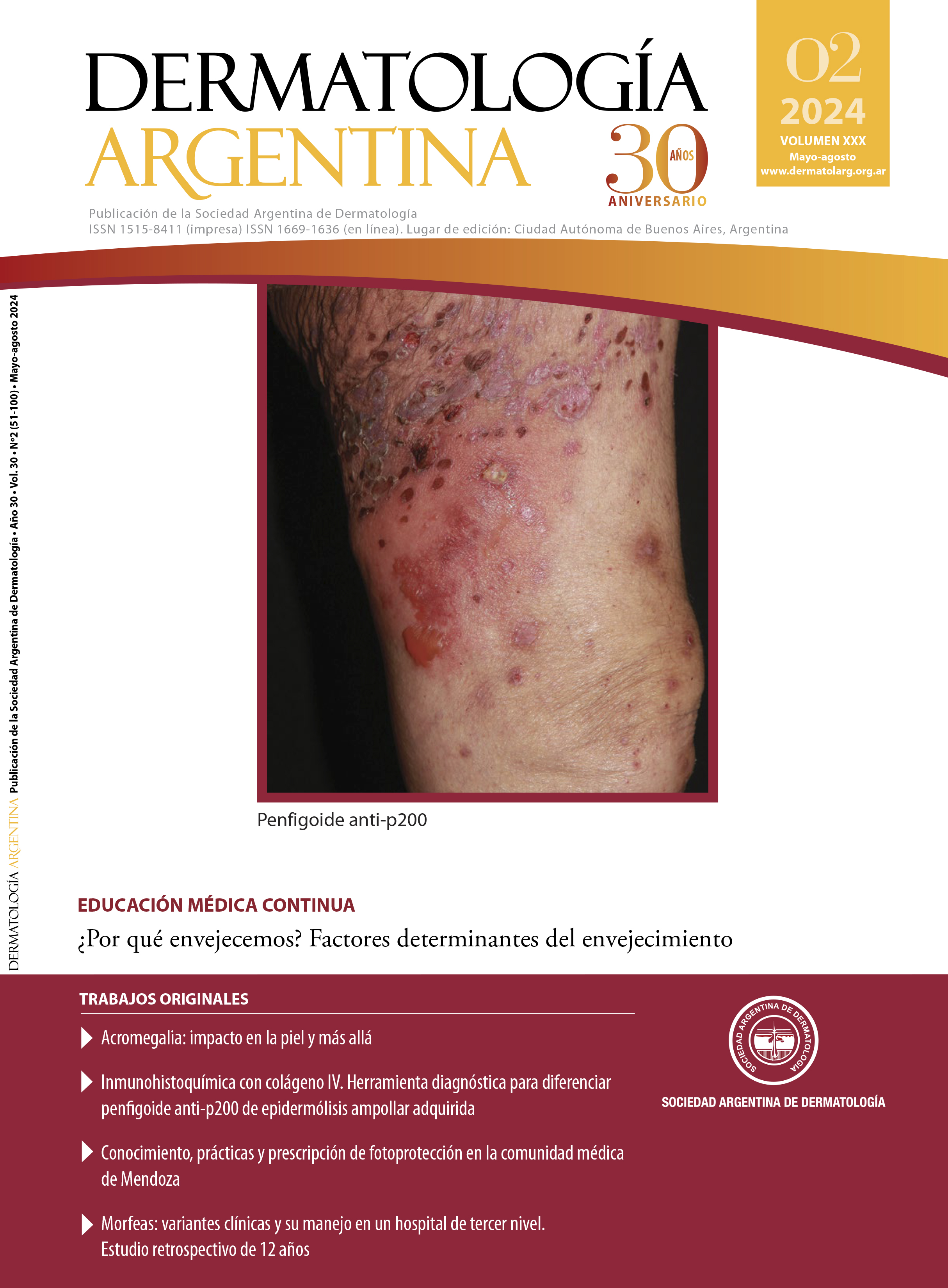
Morphea: clinical subtypes and management in a tertiary care hospital. A 12 years retrospective study
DOI:
https://doi.org/10.47196/da.v30i2.2461Keywords:
localised scleroderma, morphea, Parry Romberg syndrome, coup de sabreAbstract
Background: morphea is an inflammatory disease that primarily affects the skin and underlying tissues and results in their sclerosis. In Argentina, the statistical data on morpheas are unknown. Epidemiological studies in the United Kingdom and the United States establish that it is uncommon.
Objectives: to describe the epidemiological profile of the population diagnosed with morphea, including age, gender, types and subtypes of morphea, symptoms, associated diseases, laboratory parameters in periods of activity, and treatments performed.
Design: descriptive, observational, cross-sectional and retrospective study.
Materials and methods: analysis of medical records of patients aged 18 years or older with a diagnosis of morphea seen at the Dermatology Department of the Hospital General de Agudos José María Ramos Mejía in the Autonomous City of Buenos Aires between January 2011 and July 2023.
Results: the sample consisted of 45 patients. The median age at diagnosis was 26 years. Women accounted for 84.4%. The predominant type of morphea was linear (48.9%), followed by pansclerotic (15.6%) and circumscribed (13.3%). Within the linear type, limb morphea predominated, followed by Parry Romberg syndrome. The most frequent extracutaneous manifestation was arthralgia. Hashimoto's thyroiditis was an associated disease in 8.88% of patients. The 53.33% manifested activity parameters during follow-up. Of these, 45.8% showed elevated erythrocyte sedimentation rate. The most commonly used systemic treatment was methotrexate associated with intravenous corticosteroid pulses.
Conclusions: this is the most extensive case series of morphea published so far in Argentina. Women had a high prevalence, and the linear type was the most frequent. Although this work allows us to increase the national casuistry, the fact that the study was carried out in a third-level centre limits the extrapolation of the results to the general population of Argentina since the most severe and complex to manage cases are referred to this centre; therefore, it is essential to continue with future research.
References
I. Fett N, Werth VP. Update on morphea: Part I. Epidemiology, clinical presentation, and pathogenesis. J Am Acad Dermatol. 2011;64:217-228.
II. Silman A, Jannini S, Symmons D, Bacon P. An epidemiological study of scleroderma in the West Midlands. Br J Rheumatol. 1988;27:286-290.
III. Peterson LS, Nelson AM, Su W.P, T Mason, et ál. The epidemiology of morphea (localized scleroderma) in Olmsted County 1960-1993. J Rheumatol. 1997;24:73-80.
IV. Bielsa-Marsol I. Actualización en la clasificación y el tratamiento de la esclerodermia localizada. Actas Dermosifiliogr. 2013;104:654-666.
V. Peterson LS, Nelson AM, Su WP. Classification of morphea (localized scleroderma). May Clin Proc. 1995;70:1068-1076.
VI. Laxer RM, Zulian F. Localized scleroderma. Curr Opin Rheumatol. 2006;18:606-613.
VII. Rodríguez-Salgado P, García-Romero MT. Morfea: revisión práctica de su diagnóstico, clasificación y tratamiento. Gac Med Mex. 2019;155:522-531.
VIII. Pereira de Almeida NA, Orso-Rebellato PR, Makino Rezende Montemezzo C, Andrade-Rocha R, et ál. Morfea panesclerótica incapacitante. Dermatol Pediatr Latinoam. 2021;16(1):22-34.
IX. Ayala-Servín JN, Martínez MAD, Urizar-González CA, González M, et ál. Esclerodermia cutánea localizada (morfea): reporte de caso. Med Clin SOc. 2021;5:100-105.
X. Gaviria CM, Jiménez SB, Gutiérrez J. Morfea o esclerodermia localizada. Rev Asoc Colomb Dermatol Cir. 2014;22:126-140.
XI. Salgado PR, Zepeda CH, de Ocariz MS, Nakashimada MAY, et ál. Morphea in children: a retrospective study of its clinical characteristics and extracutaneous manifestations. Acta Pediatr Mex. 2019;40:51-58.
XII. Careta MF, Romiti R. Localized scleroderma: clinical spectrum and therapeutic update. An Bras Dermatol. 2015;90:62-73.
XIII. Pope E, Laxer RM. Diagnosis and management of morphea and lichen sclerosus and atrophicus in children. Pediatr Clin North Am. 2014;61:309-319.
XIV. Khan MA, Shaw L, Eleftheriou D, Prabhakar P, et ál. Radiologic improvement after early medical intervention in localised facial morphea. Pediatr Dermatol. 2016;33:95-98.
XV. Zwischenberger B, Jacobe HA systematic review of morphea treatments and therapeutic algorithm. J Am Acad Dermatol. 2011;65:925-941.
XVI. George R, George A, Kumar TS. Update on management of morphea (localized scleroderma) in children. Indian Dermatol Online J. 2020;11:135.
Downloads
Published
Issue
Section
License
Copyright (c) 2024 on behalf of the authors. Reproduction rights: Argentine Society of Dermatology

This work is licensed under a Creative Commons Attribution-NonCommercial-NoDerivatives 4.0 International License.
El/los autor/es tranfieren todos los derechos de autor del manuscrito arriba mencionado a Dermatología Argentina en el caso de que el trabajo sea publicado. El/los autor/es declaran que el artículo es original, que no infringe ningún derecho de propiedad intelectual u otros derechos de terceros, que no se encuentra bajo consideración de otra revista y que no ha sido previamente publicado.
Le solicitamos haga click aquí para imprimir, firmar y enviar por correo postal la transferencia de los derechos de autor


