Reglamento de publicación
Cesión de copyright
Idioma
Información
hhttps://www.dermatolarg.org.ar/
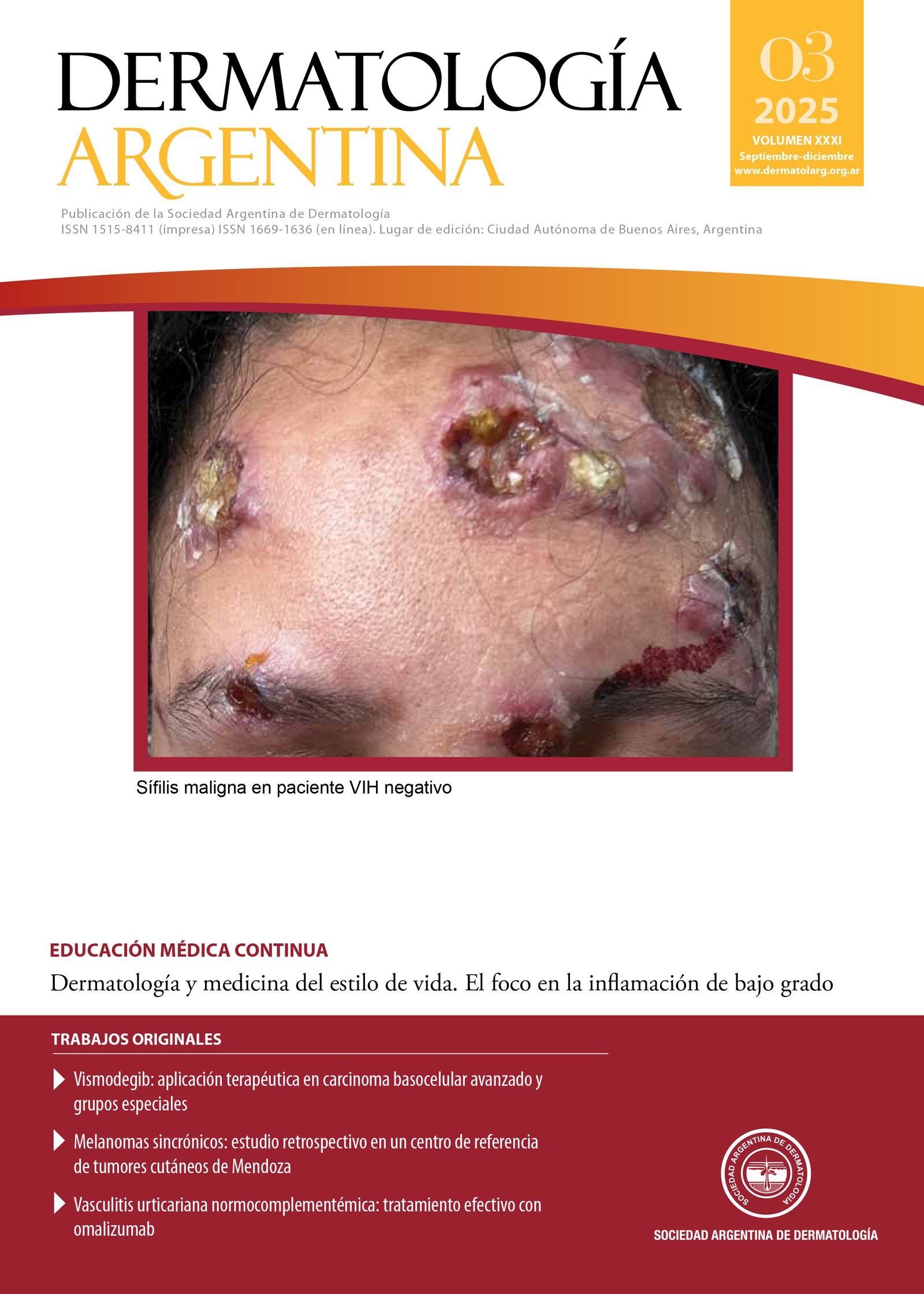
ISSN 1515-8411 (impresa) ISSN 1669-1636 (en línea)
Periodicidad cuatrimestral
Propietaria: Sociedad Argentina de Dermatología Asociación Civil (SAD)
Domicilio legal: Av. Callao 852, piso 2°, (1023), Ciudad de Buenos Aires, Argentina
Registro de la marca "Dermatología Argentina"
en Clase 9: Reg. N° 4.584.197, Acta N° 4.020.917;
en Clase 16: Reg. N° 4.584.196, Acta N° 4.050.918,
Instituto Nacional de la Propiedad Intelectual (INPI).
Registro en la Dirección Nacional de Derecho de Autor: RL-2023-71100049-APN-DNDA#MJ
Sello Editorial Lugones® de Editorial Biotecnológica S.R.L.
Av. Curapaligüe 202, piso 9°, ofic. B (1406), Ciudad de Buenos Aires, Argentina. Tel.: +54 11 4632-0701/4634-1481
administracion@lugones.com.ar | www.lugoneseditorial.com.ar


