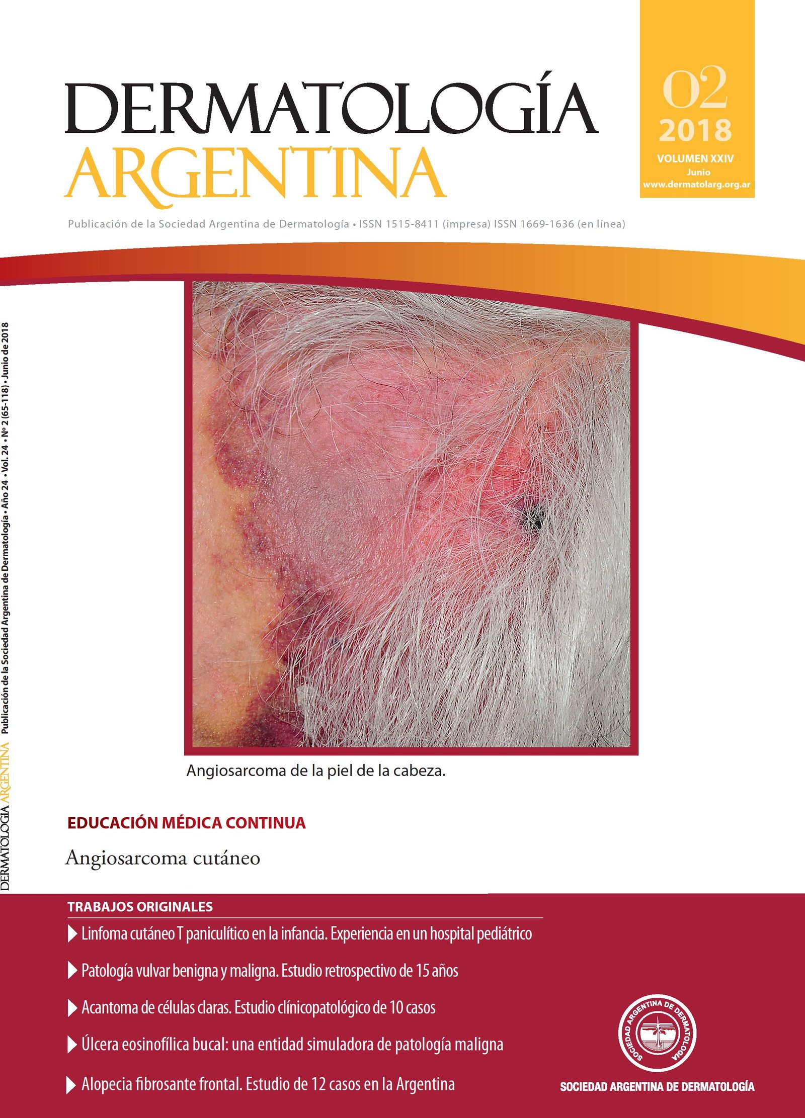
Benign and malignant vulvar pathology. A 15 year retrospective study
Keywords:
vulva, vulvar inflammatory injuries, HPV, vulvar neoplasms, precancerous conditionsAbstract
Background: Vulva can be seat of benign, malignant and non-neoplastic pathologies. Statistics in literature are limited.
Objective: To determine vulvar lesions prevalence biopsied at vulvar pathology consultation at Dermatology Unit of Hospital Lagomaggiore from Mendoza, and describe its clinical and histological characteristics.
Design: A descriptive, retrospective, observational and cross-sectional study was performed.
Materials and methods: We collected data from patients evaluated and biopsied in vulvar pathology consultation at Dermatology Unit of Hospital Lagomaggiore from Mendoza, since July 2002 to July 2017. We studied variables such as age, lesion location, evolution time, histological diagnosis and clinical-pathological correlation.
Results: A total of 2671 patients were included and 214 lesions were submitted to biopsy (8.01%). The mean age was 55.64 years (± 14.44 years). Inflammatory pathology was the most frequently (n = 72; 33.64%) and lichen sclerosus was the majority within it (n = 30; 41.67%). 90% of the patients were over 50 years old, with a higher risk of developing this condition than younger than 50 years (OR 4.37; p = 0.003). Squamous cell carcinoma (SCC) represented 75.51% (n = 37) of malignant tumors. Patients older than 50 years showed an increased risk of invasive forms (OR 1.18; p = 0.03). 28.97% (n = 62) of the patients did not know evolution time of the lesion.
Conclusions: Frequency of vulvar dermatosis observed in our hospital coincides with the literature consulted, with a significant percentage of malignant and premalignant pathologies. We emphasize the impor-tance of vulvar region examination for early diagnosis and treatment.
References
I. Heller DS. Benign tumors and tumor-like lesions of the vulva.Clin Obstet Gynecol 2015;58:526-535.
II. Mohan H, Kundu R, Arora K, Punia RS, et ál. Spectrum of vulvar lesions: a clinicopathologic study of 170 cases. Int J Reprod Contracept Obs Gynecol 2014;3:175-180.
III. Anzola GF, Lobo CJ, Márquez SM, Jurado J. Neoplasias malignas de vulva. Incidencia registrada en el servicio oncológico hospitalario de los seguros sociales. Rev Venez Oncol 2015;27:232-238.
IV. Xiao X, Meng YB, Bai P, Zou J, et ál. Vulvar cancer in China: epidemiological features
V. Renaud-Vilmer C, Dehen L, de Belilovsky C, Cavelier-Balloy B. Patología vulvar. EMC-Dermatología 2015;49:1-20.
VI. Maldonado VA. Benign vulvar tumors. Best Pract Res Clin Obstet Gynaecol 2014;28:1088-1097.
VII. Ramesh N, Anjana A, Kusum N, Kiran A, et ál. Overview of benign and malignant tumours of female genital tract. J App Pharm Sci 2013;3:140-149.
VIII. Allbritton JI. Vulvar neoplasms, benign and malignant. Obstet Gynecol Clin North Am 2017; 44:339-352.
IX. Bigby SM, Eva LJ, Leng Fong K, Jones RW. The natural history of vulvar intraepithelial neoplasia, differentiated type: evidence for progression and diagnostic challenges. Int J Gynecol Pathol 2016;35:574-584.
X. Hoang LN, Park KJ, Soslow RA, Murali R. Squamous precursor lesions of the vulva : current classification and diagnostic challenges. Pathology 2016;8:291-302.
XI. Bornstein J, Bogliatto F, Haefner HK, Stockdale CK, et ál. The 2015 International Society for the Study of Vulvovaginal Disease (ISSVD) Terminology of Vulvar Squamous Intraepithelial Lesions. Obstet Gynecol 2016;127:264-268.
XII. Halonen P, Jakobsson M, Heikinheimo O, Riska A, et ál. Lichen sclerosus and risk of cancer. Int J Cancer 2017;140:1998-2002.
XIII. Alasino M, García Llaver V, Parra V, Aredes Á, et ál. Tumores no espinocelulares de vulva. Incidencia en ocho años. Dermatol Argent 2010;17:277-283.
XIV. López N, Viayna E, San Martin M, Perulero N. Estimación de la carga epidemiológica de patologías asociadas a 9 genotipos del virus del papiloma humano en España: revisión de la literatura. Vacunas 2017;18:36-42.
Downloads
Published
Issue
Section
License
Copyright (c) 2018 Argentine Society of Dermatology

This work is licensed under a Creative Commons Attribution-NonCommercial-NoDerivatives 4.0 International License.
El/los autor/es tranfieren todos los derechos de autor del manuscrito arriba mencionado a Dermatología Argentina en el caso de que el trabajo sea publicado. El/los autor/es declaran que el artículo es original, que no infringe ningún derecho de propiedad intelectual u otros derechos de terceros, que no se encuentra bajo consideración de otra revista y que no ha sido previamente publicado.
Le solicitamos haga click aquí para imprimir, firmar y enviar por correo postal la transferencia de los derechos de autor


