Ir al contenido principal
Ir al menú de navegación principal
Ir al número actual
Ir al pie de página del sitio
ADVERTISEMENT
ADVERTISEMENT
Open Menu
Dermatología Argentina
Acerca de
Sobre la revista
Equipo Editorial
Envíos
Declaración de privacidad
Contacto
Actual
Archivos
Reglamento de publicación
Cesión de copyright
Registrarse
Entrar
Avisos
Buscar
Buscar
Registrarse
Entrar
Número actual
Vol. 31 Núm. 3 (2025): Septiembre-diciembre
DOI:
https://doi.org/10.47196/da.v31i3
Publicado:
2025-12-01
Número completo
Acceso mediante suscripción
PDF
Editorial
Compromiso y pasión
Liliana M. Olivares
111 - 111
PDF
Reflexiones de una dermatóloga feliz
Marta La Forgia
112 - 113
PDF
Educación Médica Continua
Dermatología y medicina del estilo de vida. El foco en la inflamación de bajo grado
Julieta Soledad Fischer, María Emilia Candiz, Mauro Coringrato
113 - 120
PDF
Trabajos Originales
Vismodegib: aplicación terapéutica en carcinoma basocelular avanzado y grupos especiales. Experiencia en el Hospital Prof. A. Posadas
Agustina Ragalli, Agustina María Stringa, María Laura Mauri, Roxana Di Gaeta, Roberta Pedevilla, Patricia Silvia Della Giovanna
121 - 128
PDF
Melanomas sincrónicos: estudio retrospectivo en un centro de referencia de tumores cutáneos de Mendoza
Ileana Rosalía Camardella, María del Valle Marin, María Clara Venturini, María Emilce Baiardi, Eugenia Alund, Sonia Rodríguez Saa
129 - 134
PDF
Vasculitis urticariana normocomplementémica: tratamiento efectivo con omalizumab
Ana Laura Costa, Antonella Cilio, Sabrina Merenzon, Lola Kuperman Wilder, Luciana Cabral Campana, Gabriela Bendjuia
135 - 138
PDF
Revisión
Impacto de los psicofármacos en la piel: reacciones adversas y su manejo. Revisión sistemática
Julieta Ruiz Beguerie, Berenice Fouces
139 - 145
PDF
Asociación de urticaria crónica y dermatitis atópica, un diagnóstico poco reconocido y un desafío terapéutico
María Inés Giustozzi, Ana Clara Torre, María Valeria Angles, Luis Daniel Mazzuoccolo
146 - 154
PDF
Casos Clínicos
Liquen escleroso ampollar y hemorrágico
Aldana Assad, Tatiana Delaloye, Mayled Delgado, Eliana Gerez, Gabriela Arena
155 - 157
PDF
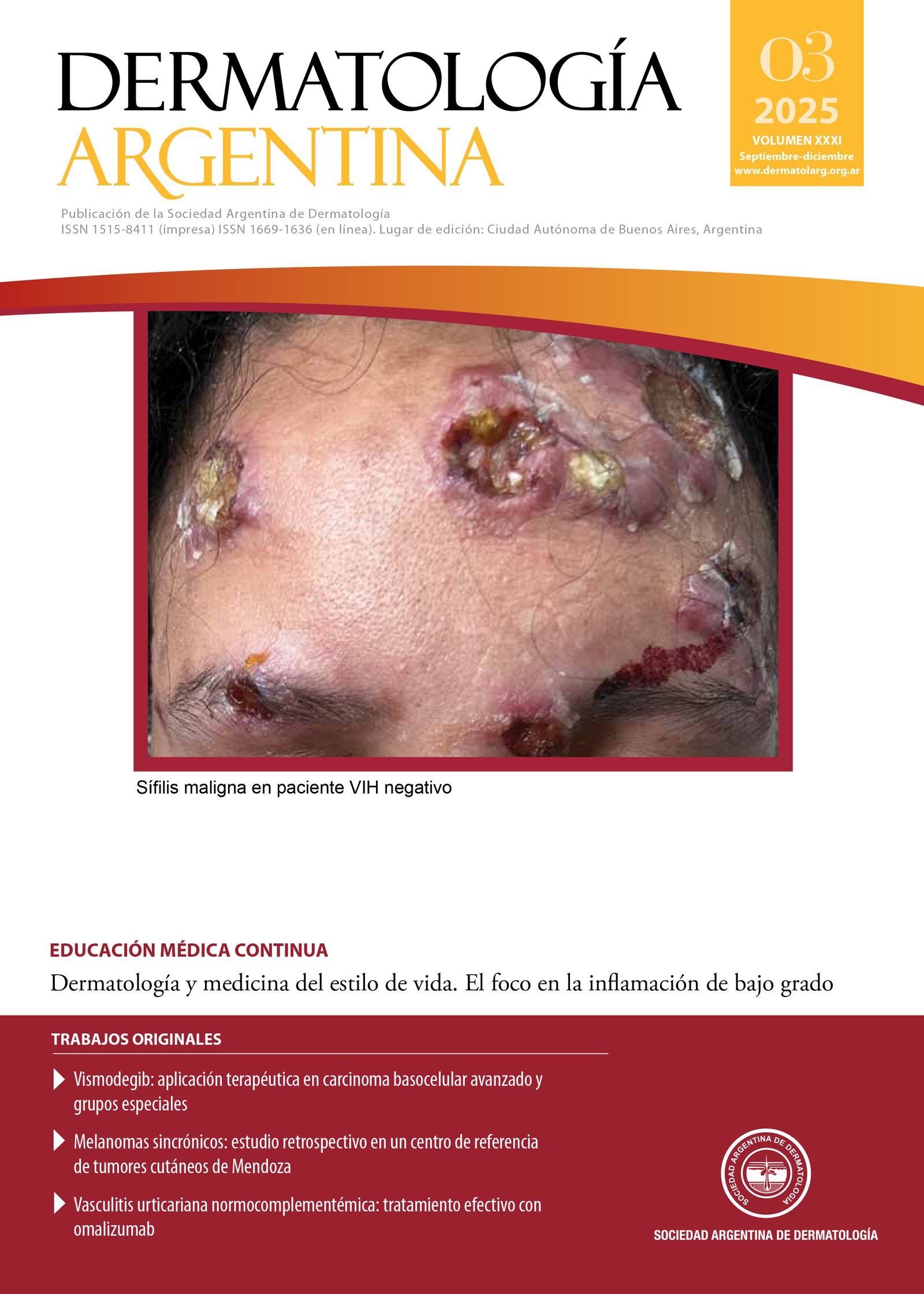
Sífilis maligna en paciente VIH negativo
María Magdalena Errandonea, Daniela Bermúdez, Andrea Torres, Julio Magliano
158 - 160
PDF
Queratodermia espinulosa adquirida
Paola Zuleta, Ornela Piñero, Mauro Coringrato, Clara Corrales
161 - 163
PDF
Psoriasis pustulosa en un paciente con astrocitoma
Pedro Mercado Puello, Yanina Berberian, Vicenta Neglia, María Inés Hernández
164 - 167
PDF
Enfermedad de Fabry: diagnóstico basado en manifestaciones cutáneas
Belén Coba, Andrea Soliani, Yanina Berberian, Mariana Demarchi, Vicenta Neglia
168 - 170
PDF
Sífilis secundaria con compromiso pulmonar
Marina Agriello, María Victoria Garritano, Paula Bonaura, Victoria Micaela Pieretti, Gabriela Laura Arena
171 - 173
PDF
Paniculitis de Weber-Christian
Agustina Alves Magalhaes, Andrea C. Soto, María L. Ortellado, María Emilia Villani
174 - 176
PDF
Carcinoma anexial microquístico de localización atípica
Camila Anderlini, Gisel Astronave, Sofía Zanitti, Marco Mazzota, Enrique Valente
177 - 179
PDF
Histoplasmosis diseminada con compromiso mucocutáneo en un paciente inmunocompetente
Candela Pagirys, Berenice Fouces, Javier Anaya, Corina Busso
180 - 182
PDF
Liquen plano hipertrófico en pediatría
María Sofía Granillo Fernández, Agostina Alonzo Caldarelli, María Victoria Moreno, Patricio Maier, Marcia Araceli Taboada
183 - 186
PDF
Dermatólogos Jóvenes
Mitos y verdades. ESCABIOSIS
Julieta Cantone, María Belén Godoy
186 - 186
PDF
¿Cuál es su Diagnóstico?
Lesiones ulceradas en miembros superiores e inferiores de 5 años de evolución
Fabiana Paola del Valle Argañaraz, Adriana Beatriz Liatto de Nogalo, Silvana Aidé López, Silvia Graciela Molina
187 - 190
PDF
Placa eritematosa en el antebrazo izquierdo
María Victoria Agüero, María Pía Herlein, Silvana Alejandra León, Graciela Luján Carabajal
191 - 192
PDF
Tumoración nodular solitaria en el antebrazo
Cynthia Laura Rossi, Leandro Danze
193 - 194
PDF
Tumoración fluctuante en la región mandibular: no solo es cuestión de piel
Carolina Belén Lorenzo, María Marta Buján, Daniel Casim, Andrea Bettina Cervini
195 - 196
PDF
La Piel en las Letras
La enfermedad y el silencio
Viviana Leiro
197 - 198
PDF
Carta al editor
Carta al editor
Carta al editor
199 - 200
PDF
Ver todos los números
ADVERTISEMENT
ADVERTISEMENT
ADVERTISEMENT
ADVERTISEMENT
ADVERTISEMENT
Idioma
English
español
Información
Para lectores/as
Para autores/as
Para bibliotecarios/as
Index-carrousel
INDEXACIÓN E INCLUSIÓN