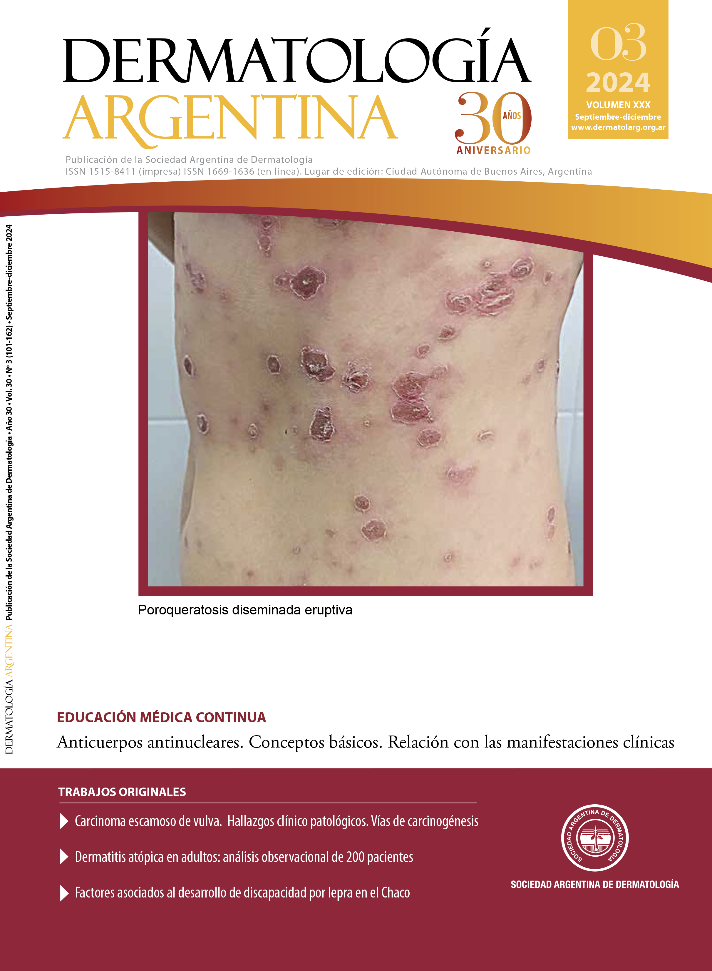
Pustules in the cervical region in a teenager
DOI:
https://doi.org/10.47196/da.v30i3.2685Keywords:
pustules, cervical region, teenagerAbstract
A 12-year-old boy consulted for a lesion located in the occipital region of the scalp that extended towards the posterior cervical region and right scapular region, which had been developing for one month. The dermatological examination revealed an erythematous plaque with multiple pustules on its Surface. At the right occipital level, a fluctuating and painful tumor was evident, with purulent secretion on the same plaque described. The pilot traction maneuver was negative. Until that moment, the patient had received multiple treatments with corticosteroids, antibiotics and topical antifungals and oral antibiotics, without response.
A direct mycological examination and culture of the lesion were requested, which was negative, so a skin biopsy of the tumor in the occipital region was performed and sent to Pathology and culture for common germs, mycobacteria and fungi. The histopathological result reported marked involvement and destruction of the hair follicle, and showed moderate edema in the papillary dermis, dense inflammatory infiltrate consisting of lymphocytes, plasma cells and numerous polymorphonuclear neutrophils and eosinophils in the papillary and reticular dermis forming foci of suppuration. No specific microorganisms were observed with the PAS technique.
References
I. Abad ME, Label A, Llorca V. Micosis superficiales. En: Larralde M, Abad E, Luna P, et ál. Dermatología Pediátrica. 3º Ed. Journal, Ciudad Autónoma de Buenos Aires, 2021: 227-232.
II. Vides De La Hoz P, Piccolomini M, Almassio A, Abad E, et ál. Tiña capitis por Trichophyton Tonsurans en un paciente pediátrico. Arch Argent Pediatr. 2022;120:e192-e196.
III. Gómez-Restrepo S, Victoria-Chaparro J. Tiña capitis en niños: pandemia aún no erradicada. Pediatr. 2022;55:142-149.
IV. Trovato MJ, Schwartz RA, Janniger CK. Tinea capitis: current concepts in clinical practice. Cutis. 2006;77:93-99.
V. Hryncewicz-Gwóźdź A, Beck-Jendroscheck V, Brasch J, Kalinowska K, et ál. Tinea Capitis and Tinea Corporis with a severe inflammatory response due to Trichophyton tonsurans. Acta Derm Venereol. 2011;91:708-710.
VI. Rodríguez A, Luna PC, Tirelli LL, Russo MF, et ál. Tiña de las barberías: una enfermedad emergente. Dermatol Argent. 2022;28:170-175.
VII. Müller VL, Kappa-Markovi K, Hyun J, Georgas D, et ál. Tinea capitis et barbae caused by Trichophyton tonsurans. A retrospective cohort study of an infection chain after shavings in barber shops. Mycoses. 2021; 64:428-436.
VIII. Gupta AK, Drummond-Main C. Meta-analysis of randomized, controlled trials comparing particular doses of griseofulvin and terbinafine for the treatment of tinea capitis. Pediatr Dermatol. 2013;30:1-6.
Downloads
Published
Issue
Section
License
Copyright (c) 2024 on behalf of the authors. Reproduction rights: Argentine Society of Dermatology

This work is licensed under a Creative Commons Attribution-NonCommercial-NoDerivatives 4.0 International License.
El/los autor/es tranfieren todos los derechos de autor del manuscrito arriba mencionado a Dermatología Argentina en el caso de que el trabajo sea publicado. El/los autor/es declaran que el artículo es original, que no infringe ningún derecho de propiedad intelectual u otros derechos de terceros, que no se encuentra bajo consideración de otra revista y que no ha sido previamente publicado.
Le solicitamos haga click aquí para imprimir, firmar y enviar por correo postal la transferencia de los derechos de autor


