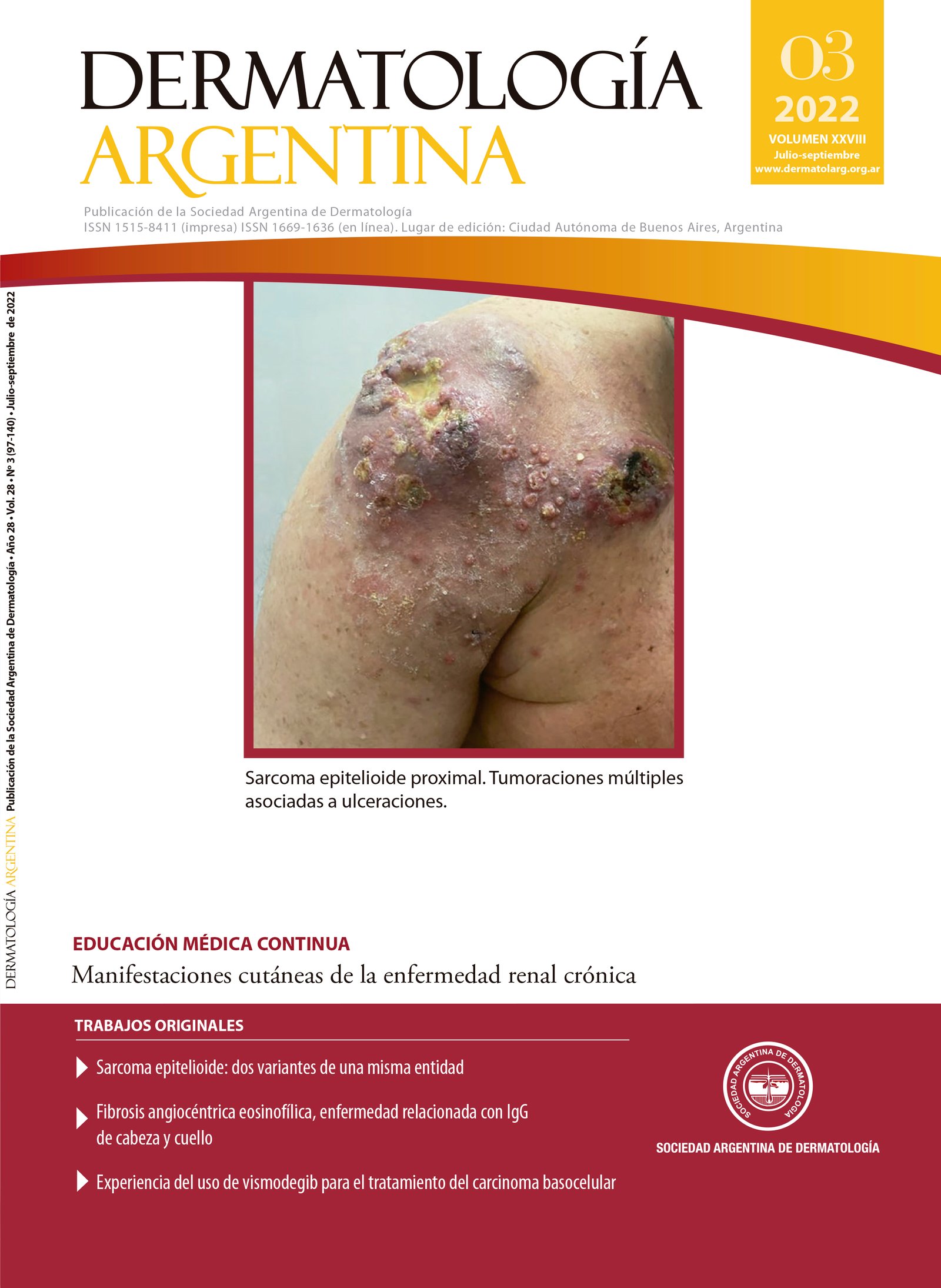
Oral superficial hemosiderotic linfovascular malformation
Keywords:
vascular malformations, oral cavity, stomatology, hemangiomaAbstract
The superficial hemosiderotic linfovascular malformation, or hobnail “hemangioma”, is an infrequent, vascular anomaly. Despite its benign nature, it requires careful diagnosis since it shares clinical and histopathological characteristics with vascular neoplasms, some of which are malignant. It exhibits a characteristic histopathological morphology, displaying cells with a “hobnail” appearance. It is generally found on the skin of the truck and extremities, rendering its location in the oral cavity extremely rare. We present the case of a patient with an oral superficial hemosiderotic linfovascular malformation of the palate.
References
I. International Society for the Study of Vascular Anomalies [internet]. ISSVA Classification of Vascular Anomalies. 2018. Disponible en: https://www.issva.org/classification. [Consultado mayo 2021]
II. Trindade F, Kutzner H, Tellechea O, Requena L, et ál. Hobnail hemangioma reclassified as superficial lymphatic malformation: A study of 52 cases. J Am Acad Dermatol. 2012;66:112-115.
III. Joyce JC, Keith PJ, Szabo S, Holland KE. Superficial hemosiderotic lymphovascular malformation (hobnail hemangioma): a report of six cases. Pediatr Dermatol. 2014;31:281-285.
IV. Mentzel T, Partanen TA, Kutzner H. Hobnail hemangioma ("targetoid hemosiderotic hemangioma"): clinicopathologic and immunohistochemical analysis of 62 cases. J Cutan Pathol. 1999;26:279-286.
V. Hejnold M, Dyduch G, Mojsa I, Okoń K. Hobnail hemangioma. Pol J Pathol. 2012;63:138-141.
VI. Alhassani M, Santhanam V, Basyuni S. Oral superficial haemosiderotic lymphovascular malformation: a rare presentation. BMJ Case Rep. 2018;2018:bcr2017223043.
VII. Yoon SY, Kwon HH, Jeon HC, Lee JH, et ál. Congenital and multiple hobnail hemangiomas. Ann Dermatol. 2011;23:539-543.
VIII. Padilla-España L, Hernández-Ibáñez C, Fúnez-Liébana R. Ecchymotic macule on a pigmented lesion following trauma. Actas Dermosifiliogr. 2014;105:707-708.
IX. 9 Hejnold M, Dyduch G, Mojsa I, Okoń K. Hobnail hemangioma: a immunohistochemical study and literature review. Pol J Pathol. 2012;63:189-192.
X. Sánchez GF, Calb IL. Hemangioma elastótico adquirido. Dermatol Argent. 2013;19:305-307.
Downloads
Published
Issue
Section
License
Copyright (c) 2022 on behalf of the authors. Reproduction rights: Argentine Society of Dermatology.

This work is licensed under a Creative Commons Attribution-NonCommercial-NoDerivatives 4.0 International License.
El/los autor/es tranfieren todos los derechos de autor del manuscrito arriba mencionado a Dermatología Argentina en el caso de que el trabajo sea publicado. El/los autor/es declaran que el artículo es original, que no infringe ningún derecho de propiedad intelectual u otros derechos de terceros, que no se encuentra bajo consideración de otra revista y que no ha sido previamente publicado.
Le solicitamos haga click aquí para imprimir, firmar y enviar por correo postal la transferencia de los derechos de autor


