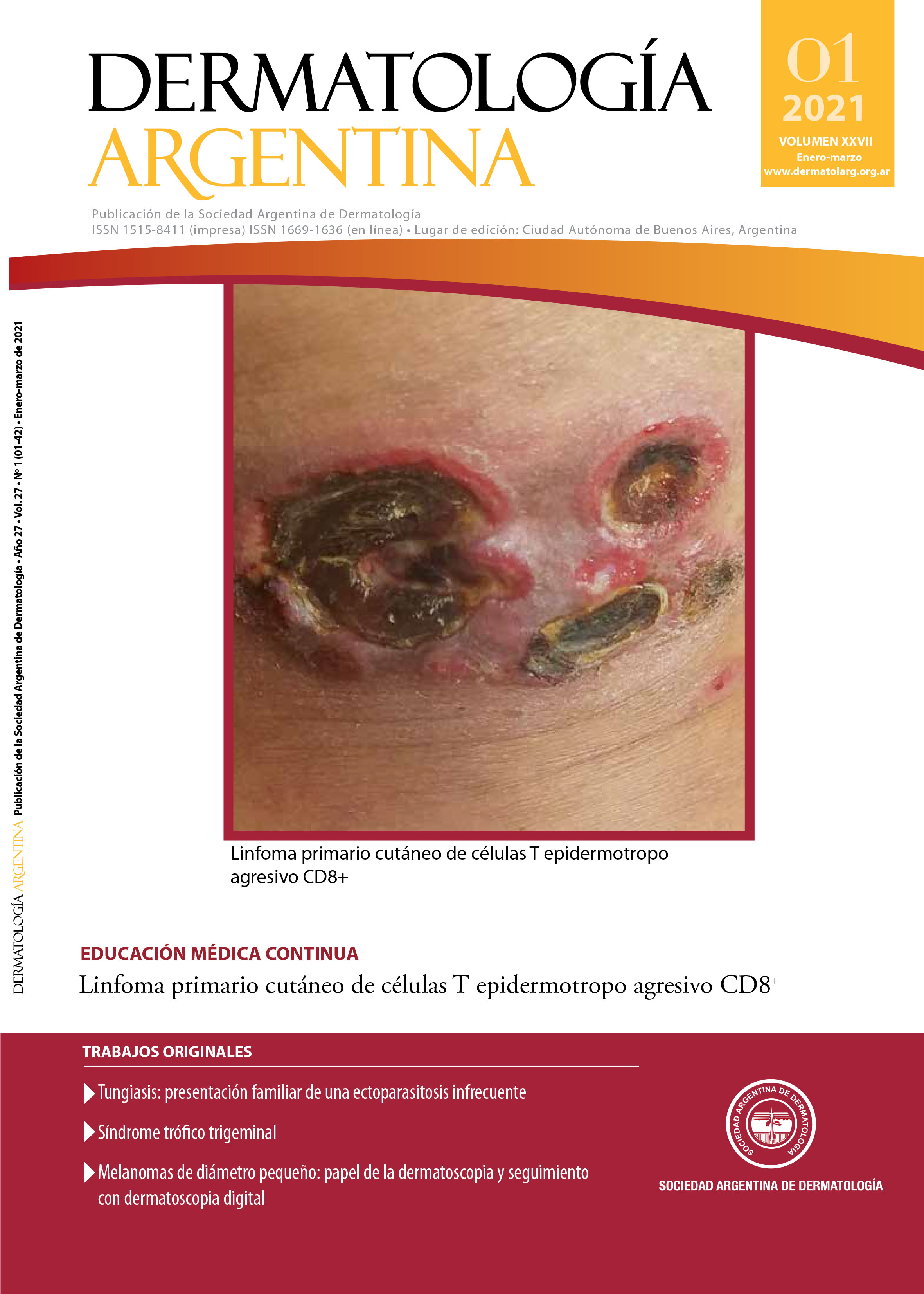
Small-diameter melanomas: role of dermoscopy and digital dermoscopy follow-up
DOI:
https://doi.org/10.47196/da.v25i1.2119Keywords:
melanoma, diagnosis, dermoscopyAbstract
Background: Recognition of small-diameter melanomas (SDM) is often challenging
Objective: To analyze the role of dermoscopy and follow-up with digital dermoscopy in the diagnosis of SDM
Design: Retrospective, observational, descriptive analysis of a series of 50 SDM diagnosed between 2015 and 2019.
Results: Among the SMD, 9 were reason for patient consultation (MMC), 30 were detected during routine nevi control (MMCR) and 11 were diagnosed due to changes observed during follow-up with digital dermoscopy (MMDD). Near 45% of the MMC were correctly classified as malignant lesions according to the “ABCD rule of dermoscopy”; this was observed only in 20% of the MMCR and in none of the MMDD. The "chaos and clues" algorithm led to excision in almost 90% of MMC, 60% of MMCR, and 50% of MMDD. The percentages of in situ melanoma were: 55% in the MMC, 73.3% in the MMCR and 72.9% in the MMDD. Among invasive melanomas, mean Breslow thickness was 0.62 mm in the MMC group, 0.5 mm in the MMCCR, and 0.4 mm in the MMDD.
Conclusions: The routine use of dermoscopy allows for the detection of melanomas with a low index of suspicion that patients may not be aware of. The use of digital follow-up enables the detection of incipient melanomas that lack not only clinical but also dermoscopic criteria of malignancy.
References
I. Kopf AW, Welkovich B, Frankel RE, Stoppelmann EJ, et ál. Thickness of malignant melanoma: global analysis of related factors. J Dermatol Surg Oncol 1987;13:345-90:401-420.
II. Argenziano G, Soyer HP, Chimenti S, Talamini R, et ál. Dermoscopy of pigmented skin lesions: results of a consensus meeting via the Internet. J Am Acad Dermatol 2003;48:679-693.
III. Salerni G, Terán T, Puig S, Malvehy J, et ál. Meta-analysis of digital dermoscopy follow-up of melanocytic skin lesions: a study on behalf of the international dermoscopy society. J Eur Acad Dermatol Venereol 2013;27:805-814.
IV. Friedman RJ, Rigel DS, Kopf AW. Early detection of malignant melanoma: the role of physician examination and self-examination of the skin. CA Cancer J Clin 1985;35:130-151.
V. Nachbar F, Stolz W, Merkle T, Cognetta AB, et ál. The ABCD rule of dermatoscopy. High prospective value in the diagnosis of doubtful melanocytic skin lesions. J Am Acad Dermatol 1994;30:551-559.
VI. Rosendahl C, Cameron A, McColl I, Wilkinson D. Dermatoscopy in routine practice-‘chaos and clues’. Aust Fam Physician 2012;41:482-487.
VII. Salerni G, Alonso C, Fernández-Bussy R. A series of small-diameter melanomas on the legs: dermoscopic clues for early recognition. Dermatol Pract Concept 2015;31;5:31-36.
VIII. Megaris A, Lallas A, Bagolini LP, Balais G, et ál. Dermoscopy features of melanomas with a diameter up to 5 mm (micromelanomas): A retrospective study. J Am Acad Dermatol 2020;83:1160-1161.
IX. Salerni G, Terán T, Alonso C, Fernández-Bussy R. The role of dermoscopy and digital dermoscopy follow-up in the clinical diagnosis of melanoma: clinical and dermoscopic features of 99 consecutive primary melanomas. Dermatol Pract Concept 2014;31;4:39-46.
Downloads
Published
Issue
Section
License
El/los autor/es tranfieren todos los derechos de autor del manuscrito arriba mencionado a Dermatología Argentina en el caso de que el trabajo sea publicado. El/los autor/es declaran que el artículo es original, que no infringe ningún derecho de propiedad intelectual u otros derechos de terceros, que no se encuentra bajo consideración de otra revista y que no ha sido previamente publicado.
Le solicitamos haga click aquí para imprimir, firmar y enviar por correo postal la transferencia de los derechos de autor


