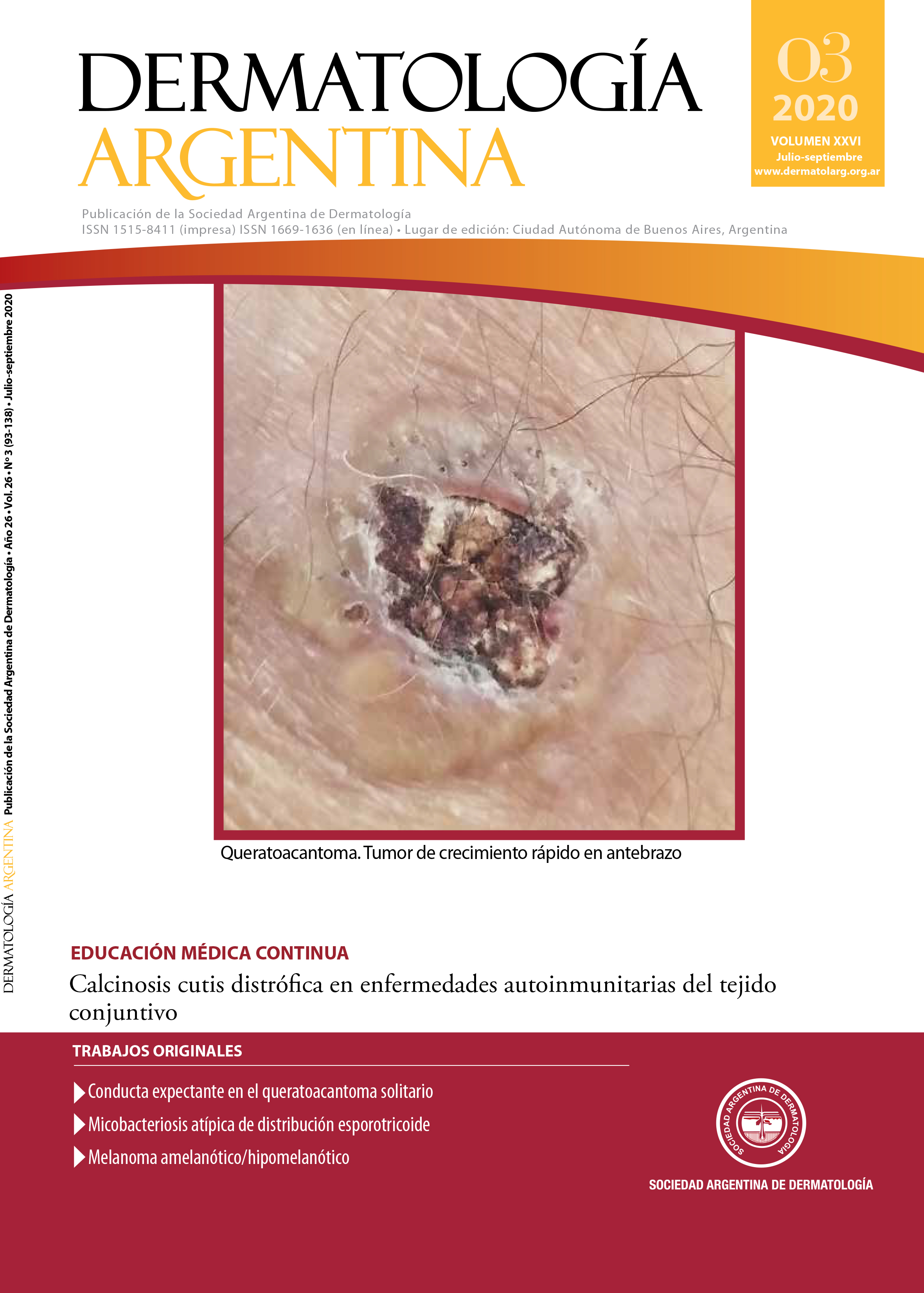
Amelanotic/hypomelanotic melanoma
DOI:
https://doi.org/10.47196/da.v26i3.2099Keywords:
amelanotic/hypomelanotic melanomaAbstract
The amelanotic/hypomelanotic melanoma is the one characterized by having a null pigment on a clinic or dermoscopic basis. It may occur in any clinic variety of melanoma. Because of its unusual characteristics, its diagnosis can take longer than expected, which entails a worse prognosis. It is found as a tumor with a taint appearance, erythematous plaque papule/nodule with scarce or null pigment, mostly found in people older than 50. In histopathology, there are no remarkable findings which allow to differentiate an hypomelanotic from any other melanoma, reason why the term should be referred to describe a clinic/dermoscopic characteristic from a determined case. Its hypomelanotic nature does not change its therapeutic behavior, which should be adjusted to the recommendations of any other melanoma, according to the stadium. We show three patients with this variety.
References
I. Rayner JE, McMeniman EK, Duffy DL, De'Ambrosis B, et ál. Phenotypic and genotypic analysis of amelanotic and hypomelanotic melanoma patients. J Eur Acad Dermatol Venereol 2019;33:1076-1083.
II. Kuznitzky R, Garay I, Kurpis M, Ruiz Lascano A. Melanoma amelanótico. Med Cutan Iber Lat Am 2003;31:202-205.
III. Gong HZ, Zheng HY, Li J. Amelanotic melanoma. Melanoma Res 2019;29:221-230.
IV. Moshe M, Levi A, Ad-El D, Ben-Amitai D, et ál. Malignant melanoma clinically mimicking pyogenic granuloma: comparison of clinical evaluation and histopathology. Melanoma Res 2018;28:363-367.
V. Ferrari A, Bono A, Baldi M, Collini P, et ál. ¿El melanoma se comporta mejor en los niños pequeños que en los adultos? Un estudio retrospectivo de 33 casos de melanoma infantil en una única institución. Pediatrics (ed. esp.). 2005;59:147-152.
VI. Zellman G. Amelanotic melanoma in a black man. J Am Acad Dermatol 1997;37:665-666.
VII. García A, Ramos L, Osorio MJ, Campos Carles A. Melanoma lentigo maligno amelanótico [en línea]. Rev Argent Dermatol 2019;100(1). Disponible en: <https://rad-online.org.ar/2019/03/31/melanoma-lentigo-maligno-amelanotico/> [Consultado marzo 2020].
VIII. Meyle KD, Guldberg P. Genetic risk factors for melanoma. Hum Genet 2009;126:499-510.
IX. Chacón M, Pfluger Y, Angel M, Waisberg F, et ál. Uncommon subtypes of malignant melanomas: a review based on clinical and molecular perspectives. Cancers 2020;2362:1-32.
X. Pouryazdanparast P, Brenner A, Haghighat Z, Guitart J, et ál. The role of 8q24 copy number gains and c-MYC expression in amelanotic cutaneous melanoma. Mod Pathol 2012;25:1221-1226.
XI. Marcucci C, Friedman P, Cesaroni E, Cohen Sabban E, et ál. Dermatoscopia del melanoma hipomelanótico y amelanótico: claves para el diagnóstico. Arch Argent Dermatol 2014;64:98-100.
XII. Detrixhe A, Libon F, Mansuy M, Nikkels-Tassoudji N, et ál. Melanoma masquerading as nonmelanocytic lesions. Melanoma Res 2016;26:631-634.
XIII. Molgó Novell M, Reyes Baraona F, Sazunic Yáñez I. Melanoma amelanótico en una paciente con enfermedad de Parkinson. Dermatol Rev Mex 2013;57:202-205.
Downloads
Published
Issue
Section
License
El/los autor/es tranfieren todos los derechos de autor del manuscrito arriba mencionado a Dermatología Argentina en el caso de que el trabajo sea publicado. El/los autor/es declaran que el artículo es original, que no infringe ningún derecho de propiedad intelectual u otros derechos de terceros, que no se encuentra bajo consideración de otra revista y que no ha sido previamente publicado.
Le solicitamos haga click aquí para imprimir, firmar y enviar por correo postal la transferencia de los derechos de autor


