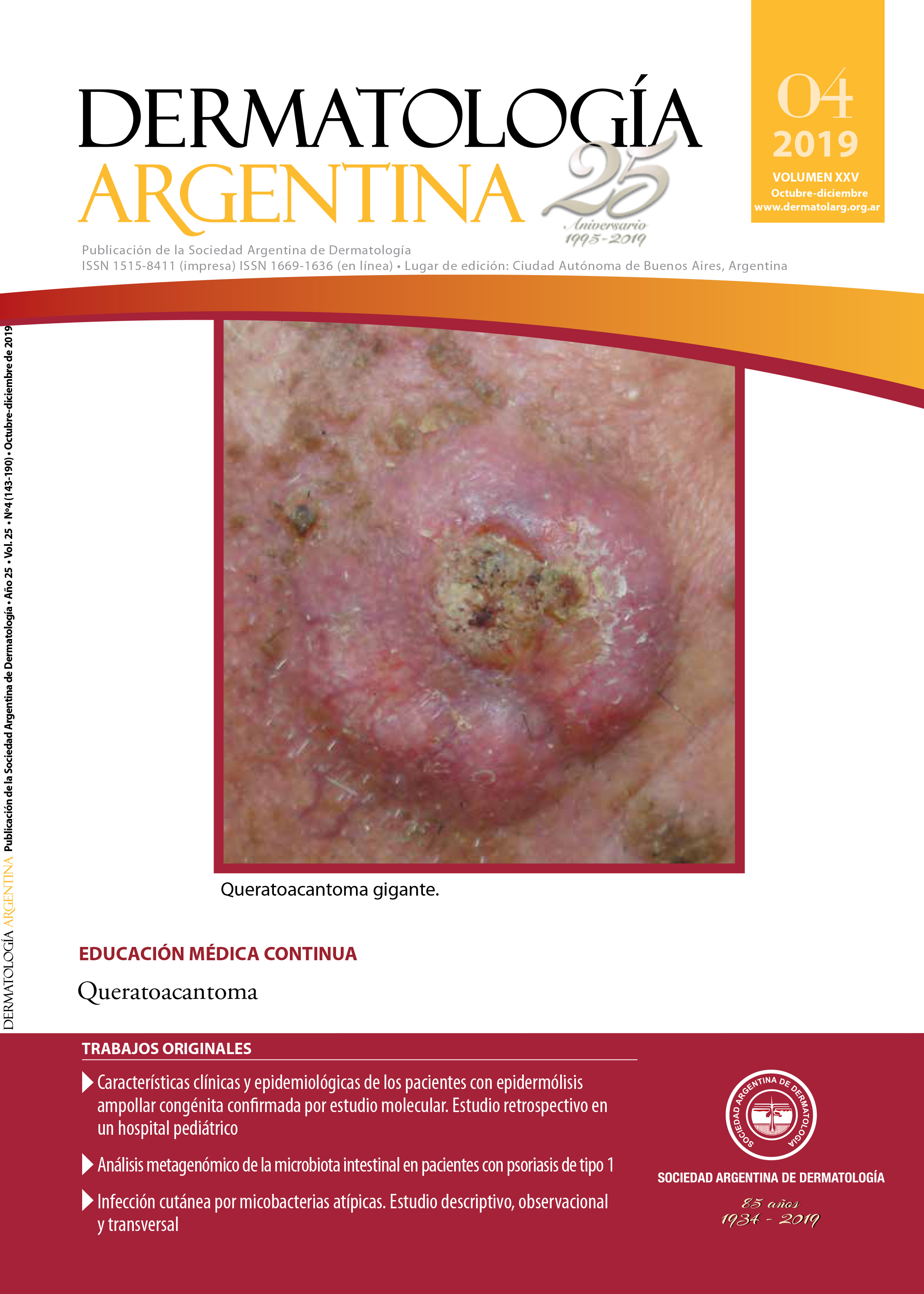
Infección cutánea por micobacterias atípicas. Estudio descriptivo, observacional y transversal
Palabras clave:
micobacterias atípicas, micobacterias no tuberculosas, micobacteriosisResumen
Antecedentes: Las micobacterias atípicas o micobacterias no tuberculosas son bacilos grampositivos, ácido-alcohol resistentes y aerobios, distribuidos amplia-mente en la naturaleza. Si bien su hallazgo ha ido en aumento, no se dispone de datos precisos, dado que no causan enfermedades de declaración obligatoria.
Objetivos: Describir la epidemiología, las manifestaciones clínicas, la histopatología y los agentes causales de las infecciones cutáneas por micobacterias atípicas en los pacientes atendidos en el Hospital de Clínicas José de San Martín durante el período comprendido entre enero de 2010 y julio de 2018.
Comparar los hallazgos obtenidos entre los pacientes inmunocompetentes y los inmunosuprimidos.
Diseño: Estudio descriptivo, observacional y transversal.
Métodos: Se revisaron las historias clínicas y los tacos de las biopsias cutáneas de todos los pacientes.
Resultados: Se incluyeron 8 pacientes: 6 mujeres y 2 hombres. Cuatro eran previamente sanos y 4, inmunosuprimidos. Los inmunocompetentes presentaron lesiones aisladas consistentes en nódulos y gomas. Los inmunosuprimidos presentaron lesiones múltiples de similares características, con predominio en los miembros inferiores. El patrón de inflamación aguda con microabscedación fue el predominante en las seis muestras analizadas. El examen directo resultó positivo en cuatro de las ocho muestras. El cultivo fue positivo en los 8 casos: 3 para M. chelonae, 2 para M. fortuitum y 1 para M. abscessus, M. kansasii y M. aviumintracellulare respectivamente.
Conclusiones: Los pacientes inmunocompetentes presentaron lesiones escasas, consistentes en nódulos y gomas coincidentes con sitios de trauma previo como fuente de la infección. Los pacientes inmunosuprimidos presentaron lesiones múltiples y diseminadas, de predominio en los miembros inferiores. La fuente de infección más frecuente fue la inyección intramuscular de metotrexato. En la histología, el patrón de inflamación aguda con microabscedación fue el predominante en ambos grupos. Nuestros resultados coincidieron con la literatura médica consultada en cuanto a epidemiología y manifestaciones clínicas. Encontramos diferencias en los hallazgos histopatológicos y microbiológicos.
Citas
I. Sander M, Isaac-Renton JL, Tyrrel GJ. Cutaneous nontuberculous mycobacterial infections in Alberta, Canadá: an epidemiologic study and review. J Cutan Med Surg 2018;22:479-483.
II. Barnes A, Rojo S, Moretto H. Prevalencia de micobacteriosis y de tuberculosis en pacientes de un hospital de referencia de la provincia de Córdoba. Rev Argen Microbiol 2004;36:170-173.
III. Caminero JA. Micobacterias atípicas. BSCP Can Ped 2001;25:237-248.
IV. Montúfar F, Madrid C, Montufar M, Aguilar C, et ál. Caracterización de pacientes hospitalizados con infecciones causadas por micobacterias no tuberculosas, en un hospital de alta complejidad de Colombia. Infectiol 2014;18:135-142.
V. Faura-Berruga C, Escario-Travesedo E, Martínez-Martínez ML, López-Villaescusa M, et ál. Micobacteriosis cutáneas y ganglionares: estudio descriptivo retrospectivo. Piel 2014;29:71-78.
VI. Alcaide F, Esteban J. Infecciones cutáneas y de partes blandas por micobacterias no tuberculosas. Enferm Infecc Microbiol Clin 2010;28:46-50.
VII. Valdés F, Cid A. Micobacterias atípicas. Actas Dermosifiliogr 2004;95:331-357.
VIII. Maroñas Jiménez L, Postigo Llorente C. Micobacteriosis cutáneas: un reto diagnóstico. Más Dermatol 2013;19:5-13.
IX. Casal M, Casal M. Las micobacterias atípicas como patógenos emergentes. Enf Emerg 2000;2:220-230.
X. Rizzo Gnatta J, Sato Kurebayashi LF, Paes da Silva M. Micobacterias atípicas asociadas a la acupuntura: revisión integral. Rev Latino-Am Enfermagem 2013;21:1-10.
XI. Misch EA, Saddler C, Davis JM. Skin and soft tissue infection due to nontuberculous mycobacteria. Curr Infect Dis Rep 2018;20:1-17.
XII. Morand J, Maslin J, Darie H. Infecciones mucocutáneas por micobacterias ambientales (incluido Mycobacterium ulcerans). EMC Dermatol 2005;39:1-21.
XIII. Moreno A, Marcoval J. Infecciones por micobacterias atípicas. En: Herrera Cevallos E, Moreno Carazo A, Requena Caballero L, Rodríguez Peralto JL. Dermatopatología: Correlación clínico-patológica. Área Científica Menarini: Madrid; 2007:295-298.
XIV. Tran A, Alby K, Kerr A, Jones M, et ál. Cost savings incurred by implementation of routine microbiologic identification by matrix-assisted laser desorption/ionization–time of flight (MALDI-TOF) mass spectrometry. J Clin Microbiol 2015;53:2473-2479.
XV. Tortoli E. Clinical manifestations of nontuberculous mycobacterium infections. Clin Microbiol Infect 2009;15:906-910.
XVI. Zárate M, Romano V, Nievas J, Smayevsky J. Utilidad de la espectrometría de masas MALDI-TOF en la identificación de bacterias anaerobias. Rev Argent Microbiol 2014;46:98-102.
XVII. Mancheno-Valencia MA, Arenas-Guzmán R, Carrillo-Casas EM, Fernández-Martínez R, et ál. La infección por micobacterias no tuberculosas, una visión desde la perspectiva dermatológica. Med Cutan Iber Lat Am 2015;43:6-13.
XVIII. Johnston R. Granulomatous reaction pattern. En: Weedon D. Weedon’s Skin Pathology. Elsevier: Londres; 2012:418-420.
XIX. Schnietz R, Wilson M. Grocott methenamine silver and periodic acid-Schiff positivity in cutaneous Mycobacterium avium complex infection. J Cutan Pathol 2018;45:551-553.
XX. Li J, Beresford R, Fyfe J, Henderson C. Clinical and histopathological features of cutaneous nontuberculous mycobacterial infection: a review of 13 cases. J Cutan Pathol 2017;44:433-443.
XXI. Olivares L, Fandiño M, Fernández Pardal P, Pérez Cortiñas ME. Infección cutánea por Mycobacterium chelonae. Dermatol Argent 2011;17:446-450.
XXII. Griffith D, Aksamit T, Brown-Elliott B, Catanzaro A, et ál. An official ATS/IDSA Statement: diagnosis, treatment and prevention of nontuberculous mycobacterial diseases. Am J Respir Crit Care Med 2007;175:367-416.
XXIII. Ortegón M, Rodríguez G, Camargo D, Orozco L. Mycobacterium chelonae y Mycobacterium abscessus: patógenos emergentes. Biomédica 1996;16: 217-238.
XXIV. del Solar M, Salomón M, Bravo F, Seas C, et ál. Infección cutánea por micobacterias atípicas de crecimiento rápido (MACR) debido a mesoterapia cosmética. Reporte de casos y revisión de la aliteratura. Folia dermatol Peru 2005;16:127-135.
XXV. Hunt C, Olivares L, Jaled M, Cergneux F. Infección por Mycobacterium marinum: a propósito de tres casos. Dermatol Argent 2013;19:332-336.
XXVI. Pastor E, Andreu A, Llombart M, Chiner E. Mycobacterium fortuitum: una rara causa de infección de marcapasos. Enferm Infecc Microbiol Clin 2006;24:136-137.
XXVII. Di Martino B, Riveros R, Medina R, Knopfelmacher O. Micobacteriosis atípica por Mycobacterium chelonae en una paciente inmunodeprimida. Presentación de un caso. Rev Panam Infectol 2013;15;47-52.
XXVIII. Bello L, Vázquez P, Rodríguez I, Predreira JD. Infección diseminada por Mycobacterium abscessus en paciente infectado por el VIH. Enferm Infecc Microbiol Clin 2012;30:47-48.
XXIX. Fica A, Soto A, Dabanch J, Porte L, et ál. Micobacterias atípicas en cinco pacientes adultos sin evidencias de inmunosupresión. Construyendo una experiencia. Rev chil infectol 2015;32:80-87.
Descargas
Publicado
Número
Sección
Licencia
El/los autor/es tranfieren todos los derechos de autor del manuscrito arriba mencionado a Dermatología Argentina en el caso de que el trabajo sea publicado. El/los autor/es declaran que el artículo es original, que no infringe ningún derecho de propiedad intelectual u otros derechos de terceros, que no se encuentra bajo consideración de otra revista y que no ha sido previamente publicado.
Le solicitamos haga click aquí para imprimir, firmar y enviar por correo postal la transferencia de los derechos de autor