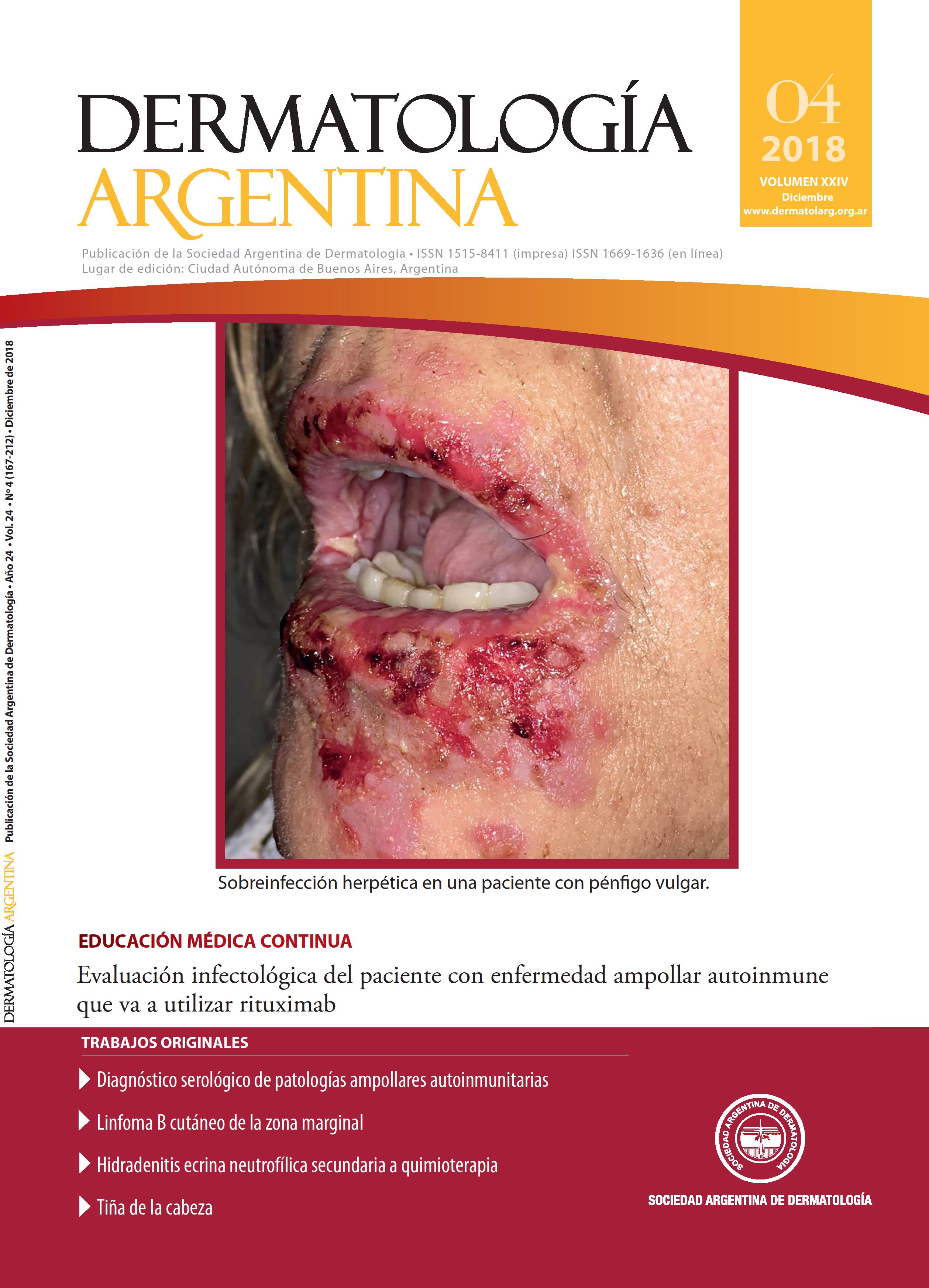
Serological diagnosis of autoimmune bullous disease
Keywords:
serology, ELISA, autoimmune bullous diseasesAbstract
Introduction: the diagnosis of autoimmune bullous disease can be a challenging task. This motivated the implementation of serology by ELISA in the Hospital de Infecciosas Dr. Francisco J. Muñiz in 2016, method unavailable in Argentina until that moment. Objectives: to communicate the results found with the implementation of this method; to correlate these results with the clinical and epidemiological characteristics of the patients; to compute the sensibility and specificity; and to make a critical analysis of this technique. Design: cross-sectional observational analytical study. Materials and methods: serological samples of patients with bullous disease from the Department of Dermatology and other health care centers were analyzed. Time period of study: from November 2016 to August 2018. Results: 63 samples were analyzed, 40 with diagnostic of pemphigus and 23 with of epidermal-dermal junction dermatoses. The sensitivity for desmoglein 1 was 87.5%, with a specificity of 100% for the diagnosis of pemphigus. As for desmoglein 3, the sensitivity was 100% and the specificity was 92.68%. In regards to BP180 and BP230, the sensitivity was 94.11% and 17.65% and the specificity was 89.13% and 95.65%, respectively. Conclusions: this is the first national study about the use of serology by ELISA for bullous diseases. The same proved to be a practical method, easy to perform, with high sensitivity and specificity for the group of pemphigus and bullous pemphigoid. On the other hand, less conclusive results were obtained in the other entities of the epidermal-dermal junction.References
I. Schmidt E, Zillikens D. The diagnosis and treatment of autoimmune blistering skin diseases. Dtsch ArzteblInt2011;108:399-405.
II. Rodríguez Costa G, Label M. Enfermedades ampollares. En: Woscoff A, Kaminsky A, Marini M, Allevato M. Dermatología en Medicina Interna. Alfaomega, Buenos Aires; 2010:147-168.
III. Rivera Díaz R, Guerra Tapia A. Novedades en dermatosis ampollares autoinmunes: pénfigos y dermatitis herpetiforme. Más Dermatol 2008;6:4-13.
IV. Pérez D, Forero O, Olivares L, Candiz ME. Dermatosis ampollares subepidérmicas neutrofílicas. Dermatol Argent2016;22:171-182.
V. Poot AM, Diercks GF, Kramer D, Schepens I, et ál. Laboratory diagnosis of paraneoplastic pemphigus. BrJ Dermatol2013;169:1016-1024.
VI. Olguín MF. Pénfigo paraneoplásico. Dermatol Argent2009;15:97-105.
VII. Campos Domínguez M, Suárez Fernández R, Lázaro Ochaita P. Métodos diagnósticos en las enfermedades ampollosas subepidérmicas autoinmunes. Actas Dermatosifiliogr2006;97:485-502.
VIII. Van Beek N, Dähnrich C, Johannsen N, Lemcke S, et ál. Prospective studies on the routine use of a novel multivariate enzyme-linked immunosorbent assay for the diagnosis of autoimmune bullous diseases. J AmAcad Dermatol2017;76:889-894.
IX. Lee EH, Kim YH, Kim S, Kim SE, et ál. Usefulness of enzymelinked immunosorbent assay using recombinant BP180 and BP230 for serodiagnosis and monitoring disease activity of bullous pemphigoid. Ann Dermatol 2012;24:45-55.
X. Le Saché-de Peufeilhoux L, Ingen-Housz-Oro S, Hue S, Sbidian E, et ál. The value of BP230 enzyme-linked immunosorbent assay in the diagnosis and immunological follow-up of bullous pemphigoid. Dermatology 2012;224:154-159.
XI. Blöcker IM, Dähnrich C, Probst C, Komorowski L, et ál. Epitope mapping of BP230 leading to a novel enzyme linked immunosorbent assay for autoantibodies in bullous pemphigoid. Br J Dermatol 2012;166:964-970.
XII. Eckardt J, Eberle FC, Ghoreschi K. Diagnostic value of autoantibody titers in patients with bullous pemphigoid. Eur J Dermatol 2018;28:3-12.
XIII. Komorowski L, Müller R, Vorobyev A, Probst C, et ál. Sensitive and specific assays for routine serological diagnosis of epidermolysis bullosa acquisita. J Am Acad Dermatol2013;68:e89-e95.
XIV. Kim JH, Kim YH, Kim S, Noh E, et ál. Serum levels of anti-type VII collagen antibodies detected by enzime-linked immunosorbent assay in patients with epidermolysis bullosa acquisita are correlated with the severity of skin lesions. J Eur Acad Dermatol Venereol 2013;27:e224-e230.
XV. Dermatology Profile ELISA (IgG).
www.euroimmun.co.uk/ukversion/img/Dermatology_Profile_ELISA.pdf.
XVI. Stanley J. Pénfigo. En: Fitzpatrick TB, Freedberg I, Eisen AZ, Wolff K, et ál.Dermatología en Medicina General. Editorial Médica Panamericana, Buenos Aires; 2005:634-645.
XVII. Ravi D, Prabhu SS, Rao R, Balachandran C, et ál. Comparison of immunofluorescence and desmoglein enzyme-linked immunosorbent assay in the diagnosis of pemphigus: a prospective, cross-sectional study in a Tertiary Care Hospital. Indian J Dermatol 2017;62:171-177.
XVIII. Lévy-Sitbon C, Reguiai Z, Durlach A, Goeldel A, et ál. Transition phénotypique d ́un pemphigus vulgaire en pemphigus superficie. Ann Dermatol Venereol 2013;140:788-792.
XIX. Marini M, Parra L, Casas J. Pénfigo herpetiforme: presentación de un caso y revisión de la literatura. Arch Argent Dermatol 2004;54:103-108
Downloads
Published
Issue
Section
License
Copyright (c) 2018 Argentine Society of Dermatolgy

This work is licensed under a Creative Commons Attribution-NonCommercial-NoDerivatives 4.0 International License.
El/los autor/es tranfieren todos los derechos de autor del manuscrito arriba mencionado a Dermatología Argentina en el caso de que el trabajo sea publicado. El/los autor/es declaran que el artículo es original, que no infringe ningún derecho de propiedad intelectual u otros derechos de terceros, que no se encuentra bajo consideración de otra revista y que no ha sido previamente publicado.
Le solicitamos haga click aquí para imprimir, firmar y enviar por correo postal la transferencia de los derechos de autor


