Ir al contenido principal
Ir al menú de navegación principal
Ir al número actual
Ir al pie de página del sitio
ADVERTISEMENT
ADVERTISEMENT
Open Menu
Dermatología Argentina
Acerca de
Sobre la revista
Equipo Editorial
Envíos
Declaración de privacidad
Contacto
Actual
Archivos
Reglamento de publicación
Cesión de copyright
Registrarse
Entrar
Avisos
Buscar
Buscar
Registrarse
Entrar
Número actual
Vol. 32 Núm. 1 (2026): Enero-abril
DOI:
https://doi.org/10.47196/ecqnsj08
Publicado:
2026-04-28
Número completo
Acceso mediante suscripción
PDF
Editorial
Salta, la ciencia nos convoca, el encuentro nos fortalece
Carolina María Ledesma
01 - 01
PDF
In memoriam
Oscar Bianchi
Comité Editorial
02 - 02
PDF
Educación Médica Continua
Trastorno por excoriación cutánea: fisiopatología y tratamiento
María Soledad Benítez, Julieta Soledad Fischer, María Emilia Candiz
03 - 11
PDF
Trabajos Originales
Análisis histopatológico de la citorreducción y del estadio 1 de slow Mohs tras electrocuretaje e imiquimod al 5% en carcinoma basocelular de alto riesgo: estudio prospectivo de 31 casos
Mauro Miguel Coringrato, Noelia Soledad García, María Emilia Candiz, María Alexandra Prado Calvo, Esteban Maronna
12 - 16
PDF
Síndrome de BASCULE en pediatría
Daniela Yoseli Puski, María Marta Bujan, Eliana Cella, Andrea Bettina Cervini
17 - 20
PDF
Prevalencia y características clínicas de farmacodermias en pacientes hospitalizados: estudio observacional transversal de 10 años en un hospital de tercer nivel en la Argentina
Mario Abbruzzese, Candela Pagirys, María Paula Isaac, Pedro Barbosa, Berenice Fouces, Corina Busso
21 - 28
PDF
Carcinoma de células de Merkel
Mary Litzi Soliz Burgos, Diego Martín Loriente, Carmen Alfaro, Patricia Silvia Della Giovanna
29 - 34
PDF
Uso de dupilumab en pacientes con dermatitis atópica y comorbilidades habitualmente excluidas de los ensayos clínicos
Macarena Nougues, Paula C. Luna, Sabrina Merenzon, Anabel Panizzardi, María Eugenia Abad, Margarita Larralde
35 - 40
PDF
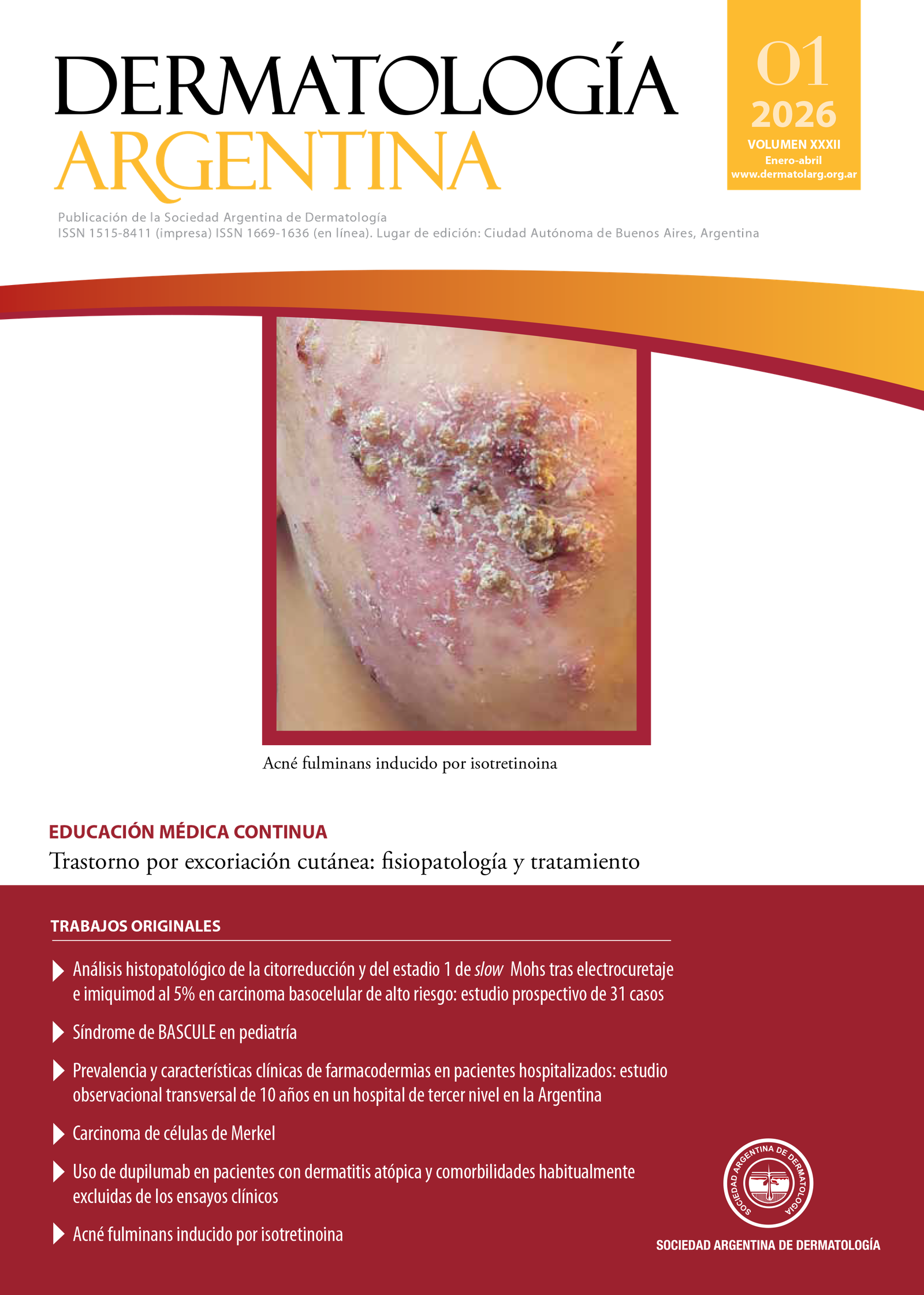
Acné fulminans inducido por isotretinoína
Felicitas Rabanal, Giselle Aignasse, Luciana Soledad Costa, Romina Foenquinos, Iris Barrio
41 - 45
PDF
Revisión
Abordaje integral de la uña en pinza: causas, diagnóstico y estrategias terapéuticas
Hilayali Aguilar Molina, Daniel Manzur Sandoval
46 - 50
PDF
Casos Clínicos
Síndrome KID con mutación p.Asp50Asn en el GJB2
Macarena Nougues, Luciana L. Tirelli, Darío Macas, John McGrath, Margarita Larralde
51 - 54
PDF
Liquen plano hipertrófico en un paciente pediátrico
Yamila Soledad Babbini, María Victoria Mayada Fabbri, María del Valle Centeno, Andrea Bettina Cervini
55 - 57
PDF
Paciente pediátrico con esporotricosis de localización inusual
Ana María Cano Valencia, Valentina Burbano Constaín, Christian Marulanda
58 - 60
PDF
Angioqueratoma circunscripto: más allá de una lesión benigna
Yesica Franco, Yanina Berberian, Andrea Soliani, María Inés Hernández, Vicenta Ana María Neglia
61 - 63
PDF
Leishmaniasis mucocutánea de aspecto lupoide
Paola Zuleta, Ludmila Rodríguez, Marisa Fernández, Clara Corrales, Viviana Leiro
64 - 66
PDF
Márgenes falsos positivos durante la cirugía micrográfica de Mohs de un carcinoma basocelular nodular en paciente con tricoepiteliomas múltiples
Ana Reggiardo, Daniela Bermúdez, Julio Magliano
67 - 69
PDF
Melanoma de la mucosa nasal: una entidad poco frecuente
Cinthia Zambrano, Silvina González, Gisella Casarotto, Horacio Solarz, Mariano Marini
70 - 73
PDF
Dermatomiositis paraneoplásica con histopatología compatible con dermatosis neutrofílica tipo Sweet
María Emilia Sívori, Isabel Hidalgo Parra, Mariana Demarchi, Rosa Beatriz Conforti, Graciela Cecilia Lozano
74 - 76
PDF
Fístula cutánea tardía secundaria a material de ligadura de safenectomía
Elena García Verdú, Cristian Fernando Caballero Linares, Paloma Gil Bernabé, José Luis Rodríguez Carrillo, Fernando Alfageme Roldán
77 - 79
PDF
Elastólisis de la dermis papilar similar a pseudoxantoma elástico
Belén Coba, María Inés Hernández, Julieta Villarroel, Leandro Danze, Vicenta Neglia
80 - 82
PDF
Dermatología Legal
El examen genital y sus implicancias médico-legales
Roberto Glorio, Sergio Carbia
83 - 85
PDF
¿Cuál es su Diagnóstico?
Dermatosis pardo-dorada
Javiela Spinelli, Melisa Giselle Baigorria, Mercedes Costantino Zanchin, María Alejandra Verea
86 - 87
PDF
Cuando lo común sorprende
María Eugenia Amoreo, Mara Lorena Ivanov, Carlos Martín, María Alejandra Verea
88 - 90
PDF
Equimosis periorbitaria
Albertina Kluver, Analía Guerra, Daniela Bermúdez, Julio Magliano
91 - 92
PDF
Lesión umbilical supurativa en paciente previamente sana
María Laura Sanz, Agustina Fernández Capiet, Alejandro Sanz, Silvana Alejandra León
93 - 94
PDF
La Piel en las Letras
Se los buscaban una a la otra
Sergio Gabriel Carbia, Roberto Glorio
95 - 96
PDF
Ver todos los números
ADVERTISEMENT
ADVERTISEMENT
ADVERTISEMENT
ADVERTISEMENT
ADVERTISEMENT
Idioma
English
español
Información
Para lectores/as
Para autores/as
Para bibliotecarios/as
Index-carrousel
INDEXACIÓN E INCLUSIÓN