Livedoid vasculopathy
Keywords:
livedoid vasculopathy, atrophie blanche, eticularis livedo, leg ulcer, blood vessel occlusion.Abstract
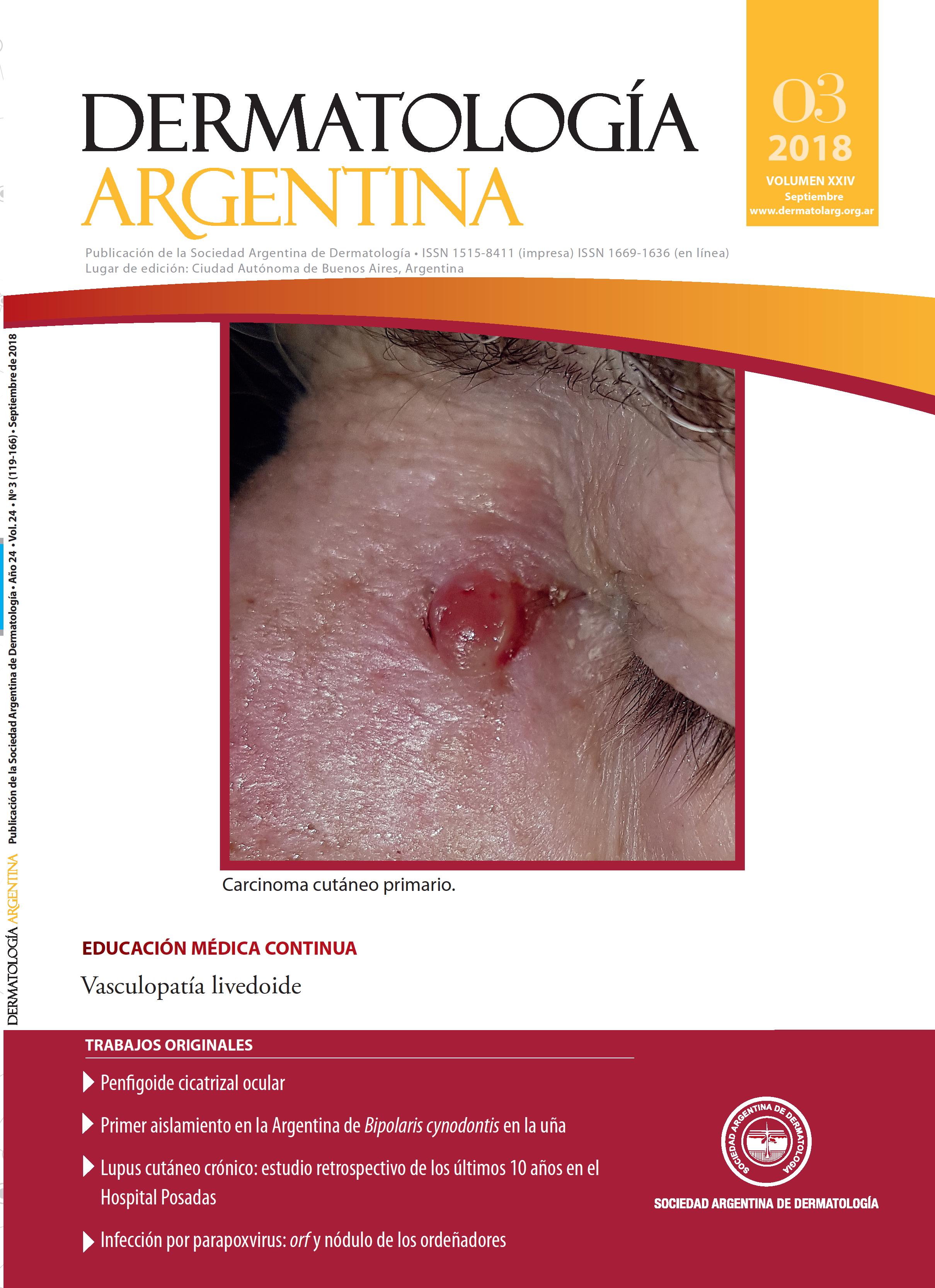
Livedoid vasculopathy (LV) is a chronic and recurrent condition caused by a state of hypercoagulability that occludes blood vessels. Its prevalence rate is 2% in the general population, and it is more frequent among young women. It usually presents painful injuries that begin as purpuric macules and erythematous papules with a livedo racemosa discoloration. These injuries evolve to gradually ulcerate and finally leave a non-pathognomonic, very characteristic pearly, star-shaped scar. Diagnosing this disease can be difficult and requires establishing a histopathological and clinical correlation. Histology varies depending on the evolutionary stage of the lesion, and the condition presents no specific signs. It is necessary to identify the underlying causes which may be triggering the fibrinolysis/thrombosis unbalance, and which are at the root of the disease. Since patients’ reactions vary greatly, there is no standardized treatment, but it must attempt to reduce pain and prevent the progression of skin lesions.
References
I. Alavi A, Hafner J, Dutz JP, Mayer D, et ál. Atrophie blanche: is it associated with venous disease or livedoid vasculopathy?Adv Skin Wound Care2014;27:518-524.
II. Papi M, Didona B, De Pità O, Frezzolini A, et ál. Livedo vasculopathy vs. small vessel cutaneous vasculitis: cytokine and platelet P-selectin studies.Arch Dermatol 1998;134:447-452.
III. Poletti ED, Sandoval NM, González JM, Torres AS. Vasculopatía livedoide: significado actual. Comunicación de dos casos. Dermatol Rev Mex 2008;52:175-181.
IV. Fernández-Antón Martínez MDC. Vasculopatía livedoide. Semin Fund Esp Reumatol 2011;12:53-56.
V. Kerk N, Goerge T. Livedoid vasculopathy-current aspects of diagnosis and treatment of cutaneous infarction.Dtsch Dermatol Ges 2013;11:407-410.
VI. Criado PR, Rivitti EA, Sotto MN, Valente NYS, et ál. Vasculopatia livedoide: uma doença cutânea intrigante. An Bras Dermatol2011;86:961-977.
VII. Tassier C, Neira MF, Anodal M, Sánchez G, et ál. Vasculopatía livedoide.Arch Argent Dermatol 2012;62:228-232.
VIII. Freitas TQ, Halpern I, Criado PR. Livedoid vasculopathy: a compelling diagnosis. Autops Case Rep 2018;8:e2018034.
IX. Feng S, Su W, Jin P, Shao C. Livedoid vasculopathy: clinical features and treatment in 24 Chinese patients.Acta Derm Venereol 2014;94:574-578.
X. Piqué E, Hernández-Machin B, Pérez-Cejudo JA, Hernández-Hernández B, et ál. Vasculopatía livedoide (atrofia blanca) generalizada en pacientes adultos con dermatomiositis.Actas Dermo-Sifiliograf 2004;95:440-443.
XI. Hu SC, Chen GS, Lin CL, Cheng YC, et ál. Dermoscopic features of livedoid vasculopathy. Medicine (Baltimore) 2017;96:e6284.
XII. Girolomoni G, Rosina P. Cutaneous vasculitides and vasculopathies. Clinical Dermatol 2013;1:93-97.
XIII. Garlatti MI, Romano MS, De los Ríos R, Saadi ME, et ál. Papulosis atrófica maligna (enfermedad de Degos).Arch Argent Dermatol 2010;60:101-104.
XIV. Hairston BR, Davis MD, Gibson LE, Drage LA. Treatment of livedoid vasculopathy with low-molecular-weight heparin: report of 2 cases. Arch Dermatol 2003;139: 987-990.
XV. Micieli R, Alavi A. Treatment for livedoid vasculopathy. A Systematic Review. JAMA Dermatol 2018;154:193-202.
XVI. Franco Marques G, Criado PR, Alvez Batista Morita TC, Cajas García MS. The management of livedoid vasculopathy focused on direct oral anticoagulants (DOACs): four case reports successfully treated with rivaroxaban. Int J Dermatol2018;57:732-741.
XVII. Jiménez-Gallo D, Villegas-Romero I, Rodríguez-Mateos ME, Linares-Barrios M. Treatment of livedoid vasculopathy with rivaroxaban: a potential use of new oral anticoagulants for dermatologists. Actas Dermosifiliogr2018;109:278-281.
XVIII. Kim EJ, Yoon SY, Park HS, Yoon HS, et ál. Pulsed intravenous immunoglobulin therapy in refractory ulcerated livedoid vasculopathy: seven cases and a literature review. Dermatol Ther 2015;28:287-290.
Downloads
Published
Issue
Section
License
Copyright (c) 2018 Argentine Society of Dermatology

This work is licensed under a Creative Commons Attribution-NonCommercial-NoDerivatives 4.0 International License.
El/los autor/es tranfieren todos los derechos de autor del manuscrito arriba mencionado a Dermatología Argentina en el caso de que el trabajo sea publicado. El/los autor/es declaran que el artículo es original, que no infringe ningún derecho de propiedad intelectual u otros derechos de terceros, que no se encuentra bajo consideración de otra revista y que no ha sido previamente publicado.
Le solicitamos haga click aquí para imprimir, firmar y enviar por correo postal la transferencia de los derechos de autor