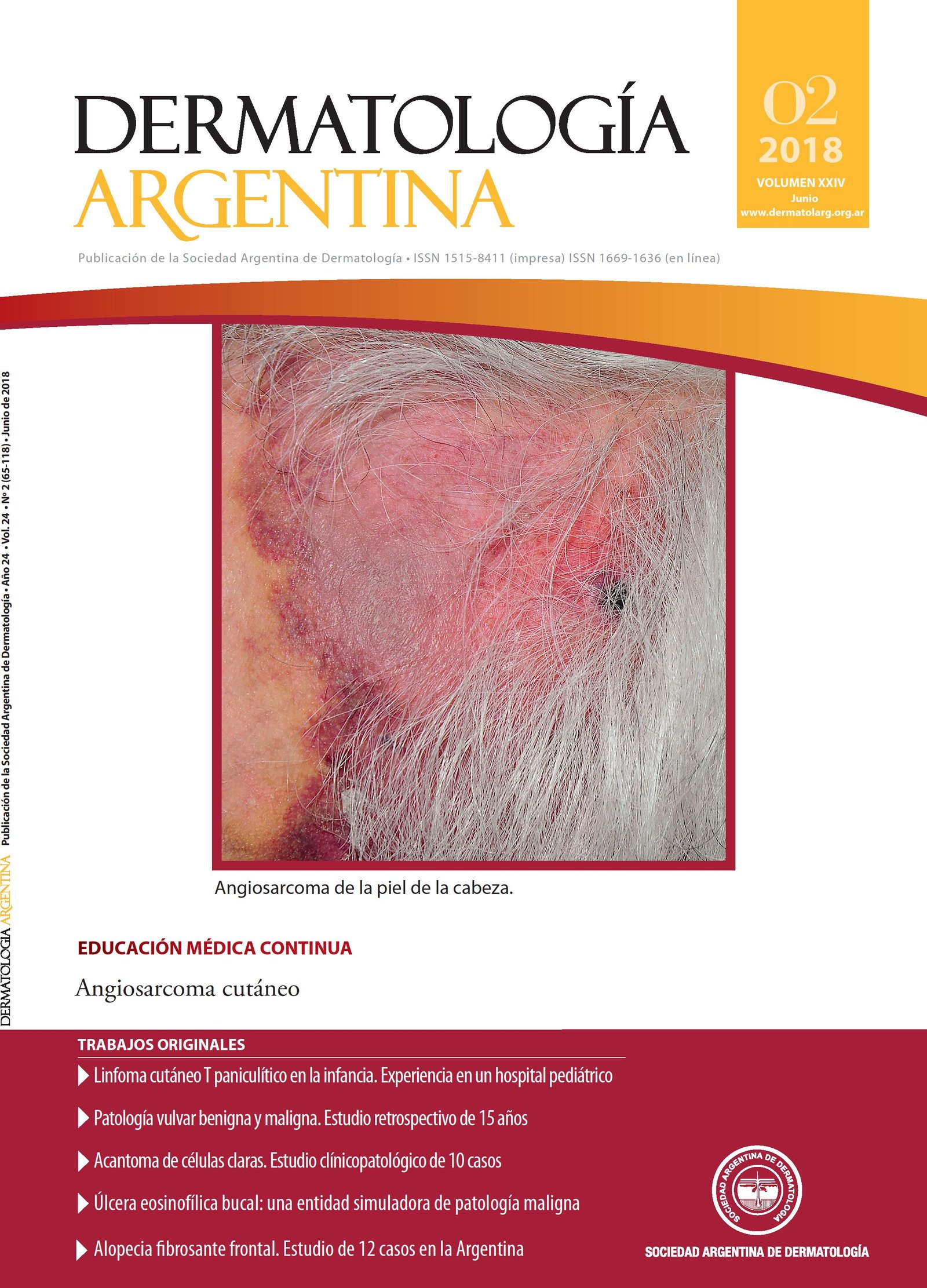
Frontal fibrosing alopecia. A study of 12 cases in Argentina
Keywords:
Frontal fibrosing alopecia, lichen planopilaris, cicatricial alopecia, PPAR, pioglitazone, dutasterideAbstract
Frontal fibrosing alopecia (FFA) is a primary scarring alopecia that typically affects postmenopausal women and is characterized by the recession of the frontal-parieto-temporal hairline with total or partial loss of eyebrows. It is an entity of chronic course with uncertain evo-lution. There are currently no internationally validated clinical-prog-nostic evaluation criteria, although a classification for this purpose has recently been proposed based on a large multicentric casuistry.
Objective: To describe the clinical and demographic characteristics of patients diagnosed with FFA assessed in two Argentine centers.
Design: Retrospective study of 12 patients with FFA evaluated be-tween August 2015 and August 2017 at the Dermatology Service of HIGA Eva Perón (San Martín, Provincia de Buenos Aires) and a private medical center (General Pico, La Pampa).
Materials and methods: The inclusion criteria was the presence of frontal-parietol-temporal recession of the hair implantation line asso-ciated to one of the following: characteristic changes of FFA (perifollicular erythema and hyperkeratosis and closure of follicular openings) and/or scalp biopsy compatible with lymphocytic scar alopecia.
Results: Twelve patients were evaluated in 2 years. All female, meno-pausal and with average age of diagnosis 69 years. Total or partial eye-brows involvement was observed in 100% of cases. The most frequent clinical pattern was the linear pattern (75%) followed by diffuse pattern (17%). All were treated with 5-alpha reductase inhibitors and top-ical minoxidil and 42% received periodic intralesional triamcinolone.
Conclusions: The cohort studied presented a mean age of diagnosis higher than the reported, but with clinical characteristics similar to those published in the international literature.
Limitations: The retrospective nature of the study and the size of the sample.
References
I. Kossard S. Postmenopausal frontal fibrosing alopecia. Scarring alopecia in a pattern distribution. Arch Dermatol 1994;30:770-774.
II. Vaño-Galvan S, Molina-Ruiz AM, Serrano-Falcón C, Arias-Santiago S, et ál. Frontal fibrosing alopecia: a multicentric review of 355 patients. J Am Acad Dermatol 2014;70:670-678.
III. Tolkachjov SN, Chaudhry HM, Camilleri MJ, Torgerson RR. Frontal fibrosing alopecia among men: A clinicopathologic study of 7 cases. J Am Acad Dermatol 2017;77:686-690.
IV. Ormaechea-Pérez N, López-Pestaña A, Subizarreta-Salvador J, Jaka-Moreno A, et ál. Alopecia frontal fibrosante en el varón: Presentación de 12 casos y revisión de la literatura. Actas Dermosifiliol 2016;107:836-844.
V. Moreno-Arrones OM, Saceda-Corralo D, Fonda-Pascual P, Rodríguez-Barata AR, et ál. Frontal fibrosing alopecia: clinical and pronostic classification. J Eur Acad Dermatol Venereol2017;31:1739-1745.
VI. Cappetta M, Hernández M, , Lucía Fiesta, Julieta Arbat, et ál. Alopecia fibrosante frontal. Dermatol Argent 2015;21:32-38.
VII. Ladizinski B, Bazakas A, Selim MA, Olsen EA. Frontal fibrosing alopecia: a retrospective review of 19 patients seen at Duke University. J Am Acad Dermatol 2013;68:749-755.
VIII. Tosti A, Miteva M, Torres F. Lonely hair: a clue to the diagnosis of frontal fibrosing alopecia. Arch Dermatol 2011;147:1240.
IX. MacDonald A, Clark C, Holmes S. Frontal fibrosing alopecia: a review of 60 cases. J Am Acad Dermatol 2012;67:955-961.
X. Pirmez R, Donati A, Valente NS, Sodré ST, et ál. Glabellar red dots in frontal fibrosing alopecia: a further clinical sign of vellus follicle involvement. Br J Dermatol 2014;170:745-746.
XI. López-Pestaña A, Tuneu A, Lobo C, Ormaechea N, et ál. Facial lesions in frontal fibrosing alopecia (FFA): Clinicopathological features in a series of 12 cases. J Am Acad Dermatol2015;73:987-992.
XII. Samrao A, Chew AL, Price V. Frontal fibrosing alopecia: a clinical review of 36 cases. Br J Dermatol 2010;163:1296-1300.
XIII. Ma SA, Imadojemu S, Beer K, Seykora JT. Inflammatory features in frontal fibrosing alopecia. J Cutan Pathol 2017;44:672-676.
XIV. Donovan JC. Finasteride-mediated hair regrowth and reversal atrophy in a patient with frontal fibrosing alopecia. JAAD Case Rep 2015;6:353-355.
XV. Mardones F, Shapiro J. Lichen planopilaris in Latin American (Chilean) population: demographics, clinical profile and treatment experience. Clin Exp Dermatol 2017;42:755-759.
XVI. Ferting R, Tosti A. Frontal fibrosing alopecia treatment options. Intractable Rare Dis Res 2016;5:314-315.
XVII. Karnik P, Tekeste Z, McCormick TS, Guilliam AC, et ál. Hair follicle stem cell-specific PPAR-gamma deletion causes scarring alopecia. J Invest Dermatol 2009;129:1243-1257.
XVIII. Ramot I, Mastrofrancesco A, Camera E, Desreumaux P, et ál. The role of PPAR g-signaling in skin biology and pathology: new targets and opportunities for clinical dermatology. Exp Dermatol 2015;24:245-251.
XIX. Mirmirani P, Karnik P. Lichen planopilaris treated with an activator-peroxisome proliferator receptor gamma agonist. Arch Dermatol 2009;145:1363-1366.
Downloads
Published
Issue
Section
License
Copyright (c) 2018 Argentine Society of Dermatology

This work is licensed under a Creative Commons Attribution-NonCommercial-NoDerivatives 4.0 International License.
El/los autor/es tranfieren todos los derechos de autor del manuscrito arriba mencionado a Dermatología Argentina en el caso de que el trabajo sea publicado. El/los autor/es declaran que el artículo es original, que no infringe ningún derecho de propiedad intelectual u otros derechos de terceros, que no se encuentra bajo consideración de otra revista y que no ha sido previamente publicado.
Le solicitamos haga click aquí para imprimir, firmar y enviar por correo postal la transferencia de los derechos de autor


