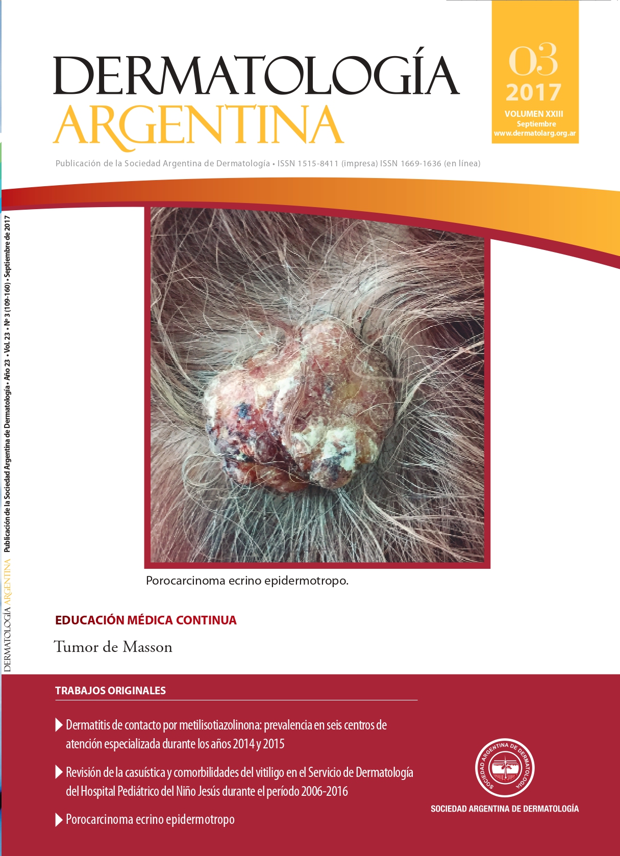
Masson ́s tumor
Keywords:
Masson tumor, reactive endothelial proliferation, benign vascular lesionAbstract
Masson’s tumor is a benign, rare vascular lesion affecting venous vessels and arteriolized venules. This reactive endothelial proliferation, which involves skin and subcutaneous tissues, is usually located in the upper and lower limbs. It presents as a palpable mass, small and superficial, redbluish. It has no predilection for age. The trau-matic factor is a frequent occurrence and the diagnosis of certainty is histopathological.The importance of knowing it is its histopathological similarity with the angiosarcoma, and the possibility of confusing the clinical diagnosis, which could lead the patient to receive unnecessary aggressive treatments.
References
I. Fernández García-Guilarte R, Enríquez de Salamanca Celada J, Comenero I. Hiperplasia papilar endotelial intravascular. Cir Plast Iberolatinoam 2009;35:155-158.
II. Fernández Figueras MT. Actualización en tumores vasculares. Hospital Universitario Germans Trias i Pujol. Universitat Autó-noma de Barcelona. http://www.conganat.org/seap/congre-sos/2003/cursodermatopatologia/: consulta 22 de diciembre de 2016.
III. Clifford PD, Temple HT, Jorda M, Marecos E. Intravascular papi-llary endothelial hyperplasia (Masson’s tumor) presenting as a triceps mass. Skeletal Radiol 2004;33:421-425.
IV. Steffen C. The man behind the eponym: CL Pierre Masson. Am J Dermatopathol 2003;25:71-76.
V. Calonje E, Wilson-Jones E. Tumores vasculares. En: Elder D, Ele-nitsas R, Jaworsky C, Johnson B. Lever. Histopatología de la piel, 8.ª ed. Buenos Aires: Intermédica, 1999:769.
VI. Weiss SW, Goldblum JR. Tumores de partes blandas. Tumores óseos de partes blandas. En: Enzinger y Weiss. Tumores benig-nos y lesiones seudotumorales de los vasos sanguíneos. Bar-celona: Elsevier España, 2009:668.
VII. Velázquez CJ, Font FI, Torres F, Araji O, et ál. Tumor de Mas-son como aneurisma de la arteria humeral. Annals Vasc Surg 2008;22:141-143.
VIII. Sangüeza O, Requena L. Cutaneous vascular hyperplasias. En: Sangüesa O, Requena L. Pathology of vascular skin lesions. Clinicopathologic Correlations. New Jersey: Humana Press, 2003:119.
IX. Akdur NC, Donmez M, Gozel S, Ustun H, et ál. Intravascular papillary endothelial hyperplasia: histomorphological and immunohistochemical features. Diagn Pathol 2013;8:167-172.
X. Ball E, Goncalves E. Hiperplasia endotelial papilar intravas-cular o pseudoangiosarcoma de Masson. Dermatol Venez2013;51:40-42.
XI. Romano MS, Gallardo C, Garlatti MI. Lesión nodular no pulsan-te color azulada. Arch Argent Dermatol 2014;64:75-76.
XII. Lee SH, Suh JS, Lim BI, Yang WI, et ál. Intravascular papillary en-dothelial hyperplasia of the extremities: MR imaging findings with pathologic correlation. Eur Radiol 2004;14:822-826.
Downloads
Published
Issue
Section
License
Copyright (c) 2017 Argentine Society of Dermatology

This work is licensed under a Creative Commons Attribution-NonCommercial-NoDerivatives 4.0 International License.
El/los autor/es tranfieren todos los derechos de autor del manuscrito arriba mencionado a Dermatología Argentina en el caso de que el trabajo sea publicado. El/los autor/es declaran que el artículo es original, que no infringe ningún derecho de propiedad intelectual u otros derechos de terceros, que no se encuentra bajo consideración de otra revista y que no ha sido previamente publicado.
Le solicitamos haga click aquí para imprimir, firmar y enviar por correo postal la transferencia de los derechos de autor


